I could be worse

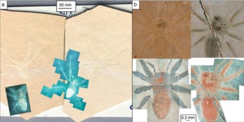
I know many of my readers shudder in dread whenever I mention “sp*d*rs”, but just imagine if Arthropleura hadn’t died off a few hundred million years ago — I’d be growing them in my lab right now and posting photos of my cuties for you to see. This is in the news now because they just found a third fossil.

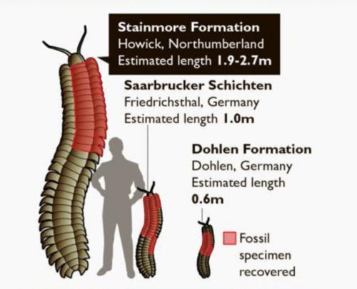
Sadly, not only have only a few of these humongous millipedes been found, but they’re all fragmentary. All we have are chunks — chunks that are several feet long — of the beast. Nobody has yet found a fossilized Arthropleura head. Just imagine all the eyes, and the nasty great mandibles, and the hungry expression on their face, if you can figure out the various bits of what passes for a face in a giant millipede. I’d show you, if I had a picture!

As long as your imagination is cranking away, here’s a visual aid.

I think his cousin is living in my bathroom shower right now.

Tell me about it

Old news.

Nothing gets between a fiercely protective mother spider and her children. Dripping tree resin trapped adult female spiders and baby spiderlings about 99 million years ago, forever showcasing the maternal care exhibited by these arthropods, according to new research.

One of the awkward things about raising spiders is that they don’t just have a few babies, and they don’t just dribble them out a few at a time over a long period…no, when spiders have babies they have a whole lot of them all at once. Yesterday, on top of all the teaching I do on Tuesdays and Thursdays, I had to feed all the spiderlings I’ve sorted out into individual vials, and then I noticed another egg sac had hatched out into a vast cloud of hungry, tiny arthropods, demanding a meal too. I’m nearly out of flies! I’m going to have to double the quantity of flies I grow just to keep up with the ravenous horde!

No one ever talks about how it was a tender parent and affectionate partner

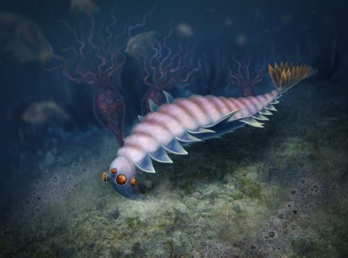
No. It’s always “underwater killing machine” this and “largest creature of its age” that. Consider the accomplishment of growing to become one of the largest animals on the planet during the Ordovician, instead.

Look at you! Scarcely out of the Cambrian, and already 2.5m long, with a sophisticated sensory system, clever system for maintaining equilibrium in the ocean, and beautifully adept tentacles. Be proud, great brave mollusc! You were more than just a murder monster.

Bring back the weird

The paleontological literature is a showcase for tragedy — it’s a graveyard of long-dead species, all snuffed out millions and millions of years before any human was around to appreciate them, and all we can do is look in awe at their fossilized corpses. In particular, fans of the Cambrian fauna can only pine for magnificently weird creatures that have been extinct for hundreds of millions of years, and represent entire exotic lineages that have left no descendants today. Two of the strangest are Anomalocaris and Opabinia.

Two of the most peculiar Burgess Shale animals, Anomalocaris and Opabinia, illustrate the complicated history of research of many Cambrian soft-bodied taxa – a result of their unfamiliar morphologies compared to the occupants of modern oceans. Both Anomalocaris and Opabinia possess compound eyes, lateral swimming flaps, filamentous setal structures, and a tail fan. Recent work has revealed that Anomalocaris and its relatives, the radiodonts, are united by the presence of paired sclerotized protocerebral frontal appendages and mouthparts composed of plates of multiple sizes, forming a diverse group containing over 20 taxa. Radiodonts range in age from the early Cambrian to at least the Devonian, and have been recovered from numerous palaeocontinents. Meanwhile, the most celebrated animal from the Burgess Shale, Opabinia regalis, with its head bearing five stalked eyes and a proboscis, remains the only opabiniid species confidently identified and is only known from a single quarry in the Burgess Shale. Myoscolex ateles from the Emu Bay Shale was proposed as a possible close relative, though this interpretation was hotly contested, and other authors have proposed a polychaete affinity.

The radiodonts (arthropods with mouths containing plates arranged in a wheel, that irised open and closed) are diverse and notorious. For a time, they were the largest predators on the planet, with their paired long spiky Great Appendages extending from the front of their head. Like the quote says, the opabiniiids are known from one location and one species, but they are weird. A similar array of swimming flaps like Anomalocaris, but then having 5 eyes on stalks and a long snout with a mouth on the end of it…it’s heartbreaking that they no longer exist. Spiders are cool and all, but I’d love to have schools of anomalocariids or opabiniids swarming in our local lakes.

At least one new opabiniid species has been identified, though. This cutie:

For perspective, here’s where they fall on the phylogenetic tree.

Tardigrades and velvet worms and mantis shrimp are certainly wonderful and interesting animals, but I have to yearn to see more of that glorious radiation of interesting forms in between. All gone, though. If gods were real, they’d never have let them die off.

Crunchies vs. Squishies: ask the pterosaurs

I’m not a taxonomist; early in my career I settled on the model systems approach, which meant all the nuances of systematics disappeared for me. “That’s a zebrafish” and “that’s not a zebrafish” were all the distinctions I had to make, and zebrafish were non-native and highly inbred so I didn’t have to think much about subtle variations. There was one taxonomic boundary one of my instructors forced me to recognize: Graham Hoyle had nothing but contempt for “squishies”, as he called vertebrates like fish or mice or people, and was much more focused on the “crunchies”, insects and crustaceans and molluscs. These seemed like odd ad hoc taxonomic categories to me, I and could think of lots of exceptions where “crunchies” were pretty squishy (see witchetty grubs or slugs), and “squishies” were armored and crunchy (armadillos, any one?), and besides, as a developmental biologist, they were all squishy if you caught them young enough. But OK, if you like dividing everything into two and only two categories, go ahead.

Then today I read this paper, “Dietary diversity and evolution of the earliest flying vertebrates revealed by dental microwear texture analysis”, and saw that there was at least one practical use for the distinction. What you eat affects wear patterns on your teeth, that if you eat lots of crunchy things vs. lots of squishy gooey things, you’ll have a different pattern of dental scratches, and since teeth fossilize — unlike guts — you can get an idea of what long dead animals had for dinner. Furthermore, you can compare fossil microwear textures to the textures in extant animals, where you do know what kinds of things they eat.

This is cool — so you can estimate the range of things ancient pterosaurs ate from how their teeth were worn, whether they ate lots of soft-bodied bugs like flies, or hard-shelled crustaceans, or soft-fleshed fish, by making a fine-grained inspection of their fossilized teeth and comparing them to modern reptiles.

a–c Reptile dietary guilds; a piscivore (Gavialis gangeticus; gharial), b ‘harder’ invertebrate consumer (Crocodylus acutus; American crocodile) and c omnivore (Varanus olivaceus; Grey’s monitor lizard). d–f Pterosaurs; d Istiodactylus, e Coloborhynchus (PCA number 5) and f Austriadactylus (PCA number 2). Measured areas 146 × 110 µm in size. Topographic scale in micrometres. Skull diagrams of extant reptiles and pterosaurs not to scale.

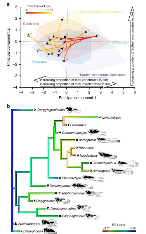
But they’re not done! Knowing the phylogenetic relationships of those pterosaurs, you can then infer evolutionary trajectories, getting an idea of how dietary preferences in species of pterosaurs shifted over time.

a Phylo-texture-dietary space of pterosaur microwear from projecting a time-calibrated, pruned tree from Lü et al.33 onto the first two PC axes of the extant reptile texture-dietary space. b Ancestral character-state reconstruction of pterosaur dietary evolution from mapping pterosaur PC 1 values onto a time-calibrated, pruned tree from Lü et al.33. To account for ontogenetic changes in diet, only the largest specimen of respective pterosaur taxa, identified by lower jaw length, were included. Pterosaur symbols same as Fig. 2. Skull diagrams of well-preserved pterosaurs not to scale (see ‘Methods’ for sources).

These results provide quantitative evidence that pterosaurs initially evolved as invertebrate consumers before expanding into piscivorous and carnivorous niches. The causes of this shift towards vertebrate-dominated diets require further investigation, but might reflect ecological interactions with other taxa that radiated through the Mesozoic. Specifically, competition with birds, which first appeared in the Upper Jurassic and diversified in the Lower Cretaceous, has been invoked to explain the decline of small-bodied pterosaurs, but this hypothesis is controversial. DMTA provides an opportunity for testing hypotheses of competitive interaction upon which resolution of this ongoing debate will depend.

In summary, our analyses provide quantitative evidence of pterosaur diets, revealing that dietary preferences ranged across consumption of invertebrates, carnivory and piscivory. This has allowed us to explicitly constrain diets for some pterosaurs, enabling more precise characterisations of pterosaurs’ roles within Mesozoic food webs and providing insight into pterosaur niche partitioning and life-histories. Our study sets a benchmark for robust interpretation of extinct reptile diets through DMTA of non-occlusal tooth surfaces and highlights the potential of the approach to enhance our understanding of ancient ecosystems.

So pterosaurs started as small bug-eaters and diversified into niches where they were consuming bigger, more diverse prey over time, which certainly sounds like a reasonable path. I don’t know that you can really assume this was a product of competition with birds — I’d want to see more info about the distribution of pterosaur species’ sizes, because expanding the morphological range doesn’t necessarily mean that you’re losing at one end of that range, but I’ll always welcome more ideas about how Mesozoic animals interacted.

Best river monster ever!

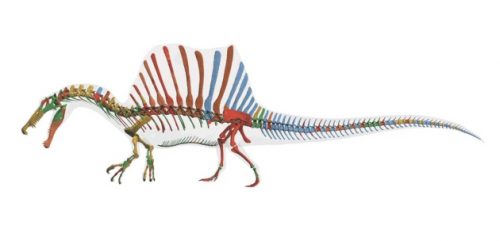
Back in 2014, a reconstruction of the full skeleton of Spinosaurus was proudly published. It had been assembled from multiple partial fossils, and was the best approximation of the organism possible.

It was an impressive beast, 15 meters long with that spectacular sail on its back.

At the time that Spinosaurus lived, what is now eastern Morocco was covered with sprawling lakes, rivers and deltas. As a top predator, the dinosaur would have had been among the rulers of an ecosystem teeming with huge crocodile-like animals, massive sawfish and coelacanths the size of cars.

Compared with other dinosaurs in its group — the two-legged, meat-eating creatures known as theropods — Spinosaurus has strikingly short rear legs. Ibrahim’s team interprets this as meaning that the dinosaur walked mainly on four legs. Its centre of gravity would have been relatively far forward, helping it to move smoothly while swimming.

John Hutchinson, a palaeontologist at the Royal Veterinary College of the University of London, is less convinced. He worries about the reliability of cobbling together different specimens to create a single picture of an animal. “We have to be careful about creating a chimera,” he says. “It’s really exciting speculation, but I’d like to see more-conclusive evidence.”

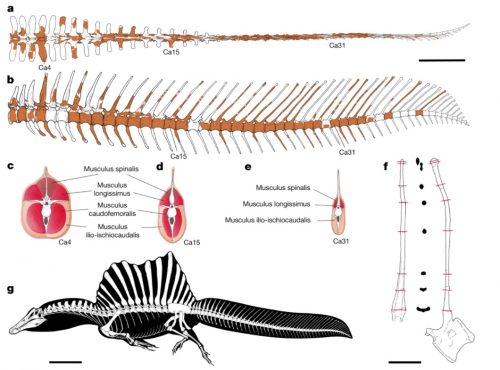
The caveat at the end was prescient. Some pieces were missing from the fossil record. Now that has been changed, and wow, it’s even more spectacular! The old tail wasn’t quite right — it had a broad paddle.

a, b, Caudal series (preserved parts shown in colour) in dorsal view (a) and left lateral view (b). c–e, Reconstructed sequential cross-sections through the tail show proximal-to-distal changes in the arrangement of major muscles. f, Sequential cross-sections through the neural spine of caudal vertebra 23 (Ca23) to show apicobasal changes. g, Skeletal reconstruction. Scale bars, 50 cm (a–e), 10 cm (f), 1 m (g).

OK, this is now my favorite dinosaur.

That is not a spider

Grrr. The CBC got me excited with a headline about “the granddaddy of spiders”. It’s not a spider. It’s a Cambrian chelicerate, which ought to be cool news enough without pretending it’s some kind of familiar organism. At least it wasn’t SciTech, which called it a frightening 500-million year old predator” or LiveScience, which called it a “nightmare creature”. C’mon, people. It was a couple of centimeters long. I do not like this pop sci nonsense that has to jack up the significance of a discovery by pretending it was scary. Does this look scary to you?

a–c, Reconstructions. a, Lateral view. b, Dorsal view (the gut has been removed for clarity). c, Isolated trunk exopod. an, anus; lam, lamellae.

At least the article by the discoverers is sensible. This is an early Cambrian chelicerate with those big old feeding appendages at the front of the head (which spiders also have) and with modified limb appendages that resemble book lungs (also a spider trait), but they are most definitely not spiders. They are their own beautiful clade, and cousins of Mollisonia plenovenatrix might have been spider ancestors, but calling them spiders is like excavating an ancient fish and calling it a mammal. Very misleading.

Yes, I’m being pedantic. It matters. Let’s not diminish the diverse chelicerates by calling them spider wanna-bes.

Here’s the abstract for the paper.

The chelicerates are a ubiquitous and speciose group of animals that has a considerable ecological effect on modern terrestrial ecosystems—notably as predators of insects and also, for instance, as decomposers. The fossil record shows that chelicerates diversified early in the marine ecosystems of the Palaeozoic era, by at least the Ordovician period. However, the timing of chelicerate origins and the type of body plan that characterized the earliest members of this group have remained controversial. Although megacheirans have previously been interpreted as chelicerate-like, and habeliidans (including Sanctacaris) have been suggested to belong to their immediate stem lineage, evidence for the specialized feeding appendages (chelicerae) that are diagnostic of the chelicerates has been lacking. Here we use exceptionally well-preserved and abundant fossil material from the middle Cambrian Burgess Shale (Marble Canyon, British Columbia, Canada) to show that Mollisonia plenovenatrix sp. nov. possessed robust but short chelicerae that were placed very anteriorly, between the eyes. This suggests that chelicerae evolved a specialized feeding function early on, possibly as a modification of short antennules. The head also encompasses a pair of large compound eyes, followed by three pairs of long, uniramous walking legs and three pairs of stout, gnathobasic masticatory appendages; this configuration links habeliidans with euchelicerates (‘true’ chelicerates, excluding the sea spiders). The trunk ends in a four-segmented pygidium and bears eleven pairs of identical limbs, each of which is composed of three broad lamellate exopod flaps, and endopods are either reduced or absent. These overlapping exopod flaps resemble euchelicerate book gills, although they lack the diagnostic operculum. In addition, the eyes of M. plenovenatrix were innervated by three optic neuropils, which strengthens the view that a complex malacostracan-like visual system might have been plesiomorphic for all crown euarthropods. These fossils thus show that chelicerates arose alongside mandibulates as benthic micropredators, at the heart of the Cambrian explosion.

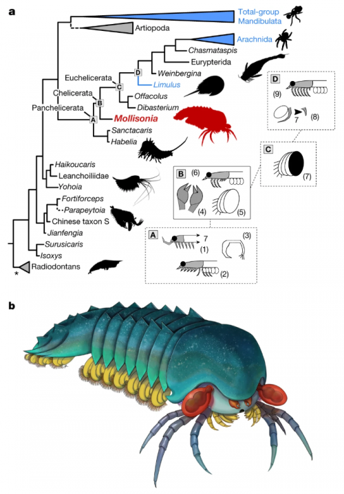
I think this diagram illustrates the relationship of M. plenoventrix to spiders well.

a, Simplified consensus tree of a Bayesian analysis of panarthropod relationships. This tree is based on a matrix of 100 taxa and 267 characters. Extant taxa are in blue; dashed branches represent questionable groupings. Asterisk shows that the radiodontans resolved as paraphyletic. This analysis excludes pycnogonids, but this had little effect on the topology. The letters A to D at the basal panchelicerate nodes refer to boxes on the right, and summarize the appearances of major morpho-anatomical features: (1) extension of cephalic shield, including a seventh tergite; (2) cephalic limbs all co-opted for raptorial and masticatory functions, and reduction of some trunk endopods; (3) dissociation of the exopod from the main limb branch; (4) presence of chelicerae; (5) trunk exopods made of several overlapping lobes; (6) some cephalic limbs differentiated as uniramous walking legs; (7) multi-lobate exopod covered by sclerite (operculum); (8) reduction of seventh cephalic appendage pair; and (9) all post-frontal cephalic limb pairs are uniramous walking legs. b, Life reconstruction. Drawing by J. Liang, copyright Royal Ontario Museum

Not a spider, but still cute and adorable.


Aria C, Caron J-B (2019) A middle Cambrian arthropod with chelicerae and proto-book gills. Nature https://doi.org/10.1038/s41586-019-1525-4.

Protists, not animals

I’ve written about the spectacular phospatized embryos of the Doushantuo formation before. It’s a collection of exceptionally well preserved small multicellular organisms, so well preserved that we can even look at cellular organelles. And they’re pre-Cambrian, as much as 630 million years old.

They’ve been interpreted as fossilized embryos for which we have no known adult forms. They certainly look like embryos, but one thing has always bothered me — they all look like blastula-stage embryos at various points in their early divisions, and the absence of later stages was peculiar: how did gastrulae and neurulae and other stages avoid getting preserved?

One explanation was that we weren’t seeing metazoan fossils at all — they were colonies of large bacteria. That’s disappointing if you have an animal bias, but still cool — as I pointed out then, it just highlights the fact that the transition from single-celled to multi-celled life isn’t that remarkable.

Now we have another alternative explanation that seems even better to me: they aren’t animals, and they aren’t bacteria, they’re protists. Some of the Doushantuo specimens are rather peanut-shaped, and others are vermiform, odd for an animal embryo, but entirely compatible with the idea that these are encysted stages of propagating protists.

Here are some of these oddly shaped Doushantuo specimens.

i-9bc3e359c5dbbed4c0f7e4e077a77157-tianzhushania.jpeg
Tianzhushania from the Ediacaran Doushantuo Formation, Datang Quarry, Weng’an, Guizhou Province, China. (A) Regular and (B to J) irregular forms, the latter interpreted to be in the germinating stage: MESIG 10022 [(A) SEM micrograph]; MESIG 10023 [(B) SEM micrograph (19)]; MESIG 10024 [(C) SEM micrograph (19)]; MESIG 10021 [(D) SEM micrograph]; SMNH X 4447 [(E) to (G) srXTM renderings]; SMNH X 4448 [(H) to (J) srXTM renderings]. (A) Surface of regular globular specimen shows envelope structure, to be compared with the similar envelope structure in (B) to (D). [(B) and (C)] Germinating specimens show protruding tubes and envelope structure. (D) Peanut-shaped specimen shows envelope structure. (E) Isosurface rendering of peanut-shaped specimen. (F) Orthoslice through (E). (G) Detail of approximate level in (F), showing cellular units. (H) Isosurface rendering of peanut-shaped specimen. (I) Orthoslice through (H). (J) Detail of approximate level in (I), showing cellular units. There is a progressive individuality of cellular units toward the periphery, including detachment of single- and oligocellular units (arrows).
i-c2ed0d62b380987fc5477a57e4735e06-lifecycle.jpeg
Proposed life cycle of Tianzhushania through hypertrophic growth of mother cell, encystment in multilayered wall, palintomic cleavage resulting in a tightly packed mass of pre-propagules, germination by opening of outer cyst wall, and release of prop- agules by degradation of inner cyst wall. Shown is the role of the outer and inner cyst walls in forming the peanut-shaped germination stages (see also modern mesomycetozoean examples in fig. S7). The outer cyst wall (seldom preserved) is indicated in black; the inner cyst wall dark is indicated in gray.

Their proposed explanation convinces me. These were protists that were single-celled in their free-living stage which would periodically grow hypertrophically and encyst, forming a capsule containing the dividing cells. These cells would replicate at differnt rates, forming zones of maturation; eventually, the cyst would rupture, released a cloud of propagules, or spores, and the life cycle would begin again.

That would explain a lot about the distribution of forms in these phosphatized specimens — we don’t find any gastrulating embryos because there never were any. These weren’t animals, period!

They belong outside crown-group Metazoa, within total-group Holozoa (the sister clade to Fungi that includes Metazoa, Choanoflagellata, and Mesomycetozoea) or perhaps on even more distant branches in the eukaryote tree. They represent an evolutionary grade in which palintomic cleavage served the function of producing propagules for dispersion.

That’s still very interesting, and again, it reminds us that the transition to multicellularity had many antecedents and could have been reached by many different paths.


Huldtgren T, Cunningham JA, Yin C, Stampanoni M, Marone F, Donoghue PC, Bengtson S (2011) Fossilized nuclei and germination structures identify Ediacaran “animal embryos” as encysting protists. Science 334(6063):1696-9.

(Also on FtB)

The eyes of Anomalocaris

Look with your puny camera eyes! Some new specimens of Anomalocaris, the spectacular Cambrian predator, have been discovered in South Australia. These fossils exhibit well-preserved eyes, allowing us to see that the bulbous stalked balls on their heads were actually fairly typical compound eyes, like those of modern insects.

i-488c66eb96bbfef0beee7fdb0bf4f590-anomalocaris_eyes-thumb-500x418-71443.jpeg

Anomalocaris eyes from the Emu Bay Shale. a-d, Eye pair, SAM P45920a, level 10.4 m. a, b, Overview and camera lucida drawing. Scale bars, 5 mm. Grey fill in b represents visual surface, the proximal part in the upper eye extrapolated from the lower eye. c, Detail of ommatidial lenses located by horizontal white box in a. Scale bar, 1 mm. d, More complete eye, showing transition between visual surface and eye stalk (white arrows). Scale bar, 2 mm. e, Detail of ommatidial lenses in counterpart SAM P45920b. Scale bar, 0.3 mm. es, eye stalk; I.c., Isoxys communis; us, undetermined structure; vs, visual surface. Tilted white box in a represents area analysed using SEM-EDS.

The cool part of this discovery: the investigators were able to count the density of lenses and estimate how many were present in the intact eye. The number is 16,000 ommatidia in each eye, which is more than a little impressive: to put it in context, Drosophila has about 800. The emphasis on high-resolution vision suggests that Anomalocaris was diurnal predator in shallow water.

Oh, and just in case you’re one of those strange beings who isn’t instantly familiar with what the anomalocarids looked like, here’s a video to remind you.


Paterson JR, García-Bellido DC, Lee MS, Brock GA, Jago JB, Edgecombe GD (2011) Acute vision in the giant Cambrian predator Anomalocaris and the origin of compound eyes. Nature 480(7376):237-40.

(Also on FtB)

Creationist abuse of cuttlefish chitin

A few weeks ago, PLoS One published a paper on the observation of preserved chitin in 34 million year old cuttlebones. Now the Institute for Creation Research has twisted the science to support their belief that the earth is less than ten thousand years old. It was all so predictable. It’s a game they play, the same game they played with the soft tissue preserved in T. rex bones. Here’s how it works.

Compare the two approaches, science vs. creationism. The creationists basically insert one falsehood, generate a ludicrous conflict, and choose the dumbest of the two alternatives.

The Scientific Approach

find traces of organic material in ancient fossils

Cool! We have evidence of ancient biochemistry!

Science!

The Creationist Approach

declare it impossible for organic material to be ancient

steal other people’s discovery of organic material in ancient fossils

Cool! Declare that organic material must not be ancient, because of step 1, which we invented

Throw out geology, chemistry, and physics because they say the material is old

Profit! Souls for my Lord Arioch!

You see, the scientists are aware of the fact that organic materials degrade over time, but recognize that we don’t always know the rate of decay under all possible conditions. When we find stuff that hasn’t rotted away or been fully replaced by minerals, we’re happy because we’ve got new information about ancient organisms, and we may also be able to figure out what mechanisms promoted the preservation of the material.

The creationists start with dogma — in this case, a false statement. They declare

Chitin is a biological material found in the cuttlebones, or internal shells, of cuttlefish. It has a maximum shelf life of thousands of years…

and

Because of observed bacterial and biochemical degradation rates, researchers shouldn’t expect to find any original chitin (or any other biomolecule) in a sample that is dozens of millions of years old–and it therefore should be utterly absent from samples deposited hundreds of millions of years ago. Thus, the chitin found in these fossils refutes their millions-of-years evolutionary interpretation, just as other fossil biomolecules already have done.

But wait. How do they know that? The paper they are citing says nothing of the kind; to the contrary, it argues that while rare, other examples of preserved chitin have been described.

Detection of chitin in fossils is not frequent. There are reports of fossil chitin in pogonophora, and in insect wings from amber. Chitin has also been reported from beetles preserved in an Oligocene lacustrine deposit of Enspel, Germany and chitin-protein signatures have been found in cuticles of Pennsylvanian scorpions and Silurian eurypterids.

So the paper is actually saying that the “maximum shelf life” of chitin is several tens of millions of years. And then they go on to describe…chitin found in Oligocene cuttlefish, several tens of millions of years old. The creationists are busily setting up an imaginary conflict in the evidence, a conflict that does not exist and is fully addressed in the paper.

The creationists do try to back up their claims, inappropriately. They cite a couple of papers on crustacean taphonomy where dead lobsters were sealed up in anoxic, water- and mud-filled jars; they decayed. Then they announce that there’s only one way for these cuttlebones to be preserved, and that was by complete mineralization, and the cuttlebones in the PLoS One paper were not mineralized.

…mineralization–where tissues are replaced by minerals–is required for tissue impressions to last millions of years. And the PLoS ONE researchers verified that their cuttlebone chitin was not mineralized.

Funny, that. You can read the paper yourself. I counted 14 uses of the words “mineralized” and “demineralized”. They state over and over that they had to specifically demineralize the specimens in hydrochloric acid to expose the imbedded chitin. And of course the chitin itself hadn’t been mineralized, or it wouldn’t be chitin anymore! Did the creationists lie, or did they just not understand the paper?

The scientists also do not claim that the chitin has not been degraded over time. They actually document some specific, general properties of decay in the specimens.

β-chitin is characterized by parallel chains of chitin molecules held together with inter-chain hydrogen bonding. The OH stretching absorbance, at about 3445 cm−1 in extant chitin, is diminished in the fossil and shifted to lower wavenumbers, showing that the specimen is losing OH by an as yet undetermined mechanism. The N-H asymmetric stretching vibration is shifted to slightly lower wavenumbers, showing that it is no longer hydrogen bonding exactly as in extant specimens. Changes in the region 2800-3600 cm−1 indicate that biomolecules have been degraded via disruption of interchain hydrogen bonds.

So, yes, the creationists seem to have rather misrepresented what the paper said. Here’s another blatant example of lying about the contents of the paper.

The question they did not answer, however, is why the original organic chitin had not completely fallen apart, which it would have if the fossils with it were 34 million years old…

Actually, they did. A substantial chunk of the discussion was specifically about that question, a consideration of the factors that contributed to the preservation. It was a combination of an anoxic environment, the presence of molecules that interfered with the enzymes that break down chitin, and the structure of cuttlebone, which interleaves layers containing chitin with layers containing pre-mineralized aragonite.

In vivo inorganic-organic structure of the cuttlebone, in combination with physical and geochemical conditions within the depositional environment and favorable taphonomic factors likely contributed to preservation of organics in M. mississippiensis. Available clays within the Yazoo Clay in conjunction with suboxic depositional environment may have facilitated preservation of original organics by forming a physical and geochemical barrier to degradation. One key to the preservation of organic tissues, in particular chitin and chitosan, is cessation of bacterial degradation within environments of deposition. Bacterial breakdown of polymeric molecules is accomplished through activities of both free extracellular enzymes (those in the water column) and ektoenzymes (those on the surface of the microbial cell) such as chitinases. Chitinases function either by cleaving glycosidic bonds that bind repeating N-acetyl-D-glucosamine units within chitin molecules or by cleaving terminal N-acetyl-D-glucosamine groups. These enzymes adsorb to the surface of clay particles, which inactivates them. Strong ions in solution like iron may act in the same manner. Once bound to functional groups within these polymeric molecules, Fe2+ ions prevent specific bond configuration on the active-site cleft of specific bacterial chitinases and prevents hydrolysis, thus contributing to preservation.

Organic layers within cuttlebones are protected by mineralized layers, similar to collagen in bones, and this mineral-organic interaction may also have played a role in their preservation. Specimens of M. mississipiensis show preserved original aragonite as well as apparent original organics. These organics appear to be endogenous and not a function of exogenous fungal or microbial activity. Fungi contain the γ form of chitin not the β allomorph found in our samples. Also, SEM analyses shows there is no evidence of tunneling, microbes, or wide-spread recrystallization of the aragonite. Therefore the chitin-like molecules detected in fossil sample are most likely endogenous. Similar to collagen in bone, perhaps, organics could not be attacked by enzymes or other molecules until some inorganic matrix had been removed.

Like I always say, never ever trust a creationists’ interpretation of a science paper: they don’t understand it, and they are always filtering it through a distorting lens of biblical nonsense. They make such egregious errors of understanding that you’re always left wondering whether they are actually that stupid, or that sleazily dishonest. Or both.

But imagine if the creationists hadn’t screwed up royally in reading the paper, if they had actually found an instance of scientists being genuinely baffled by a discovery that should not be. What if there was actually good reason to believe that chitin could not last more than ten thousand years?

Then the only sensible interpretation of this observation of 34 million year old chitin would be that the prior estimation of the shelf-life of chitin was wrong, and that it could actually last tens of millions of years. What the creationists want to do is claim that that minor hypothetical is actually correct, and that instead the entirety of nuclear physics, geology, radiochemistry, and modern cosmology is wrong. On the one hand, uncertain details about the decay of one organic molecule; in the other, entire vast fields of science, already verified, and with complex modern technologies built on their operation…and which hand would the creationists reject? The trivial one, of course.

I’m leaning towards “stupid” as the explanation for their bad arguments.


Oh, after I started this dissection, I discovered someone had already beaten me to it: here’s another analysis of the creationist misinterpretations.

(Also on FtB)