Everyone likes cute furries more than spiders, I’ve noticed

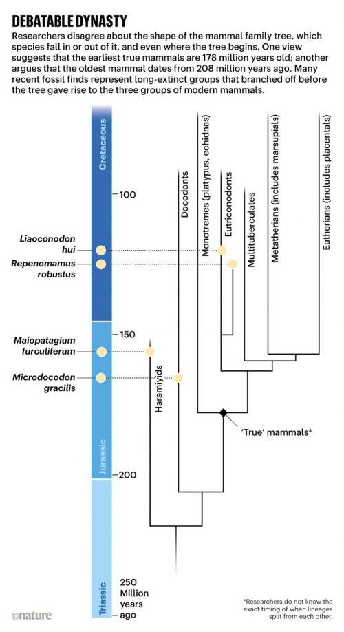
I can’t be the only one who reads outside my discipline to get material to help me cover all those evolutionary phenomena I know little about. I know a bit about fish and arthropods, but my understanding of the details of mammalian evolution is a bit thin — yet for some reason, students are more interested in the history of mammals than of spiders. I really appreciate it when I stumble across information that fills in the gaps in my knowledge in presentable ways, and Nature has done just that with a graphically rich article on How the earliest mammals thrived alongside dinosaurs. There is lots of good stuff here, and I particularly like the emphasis on the importance of fossilized infants. Development matters!

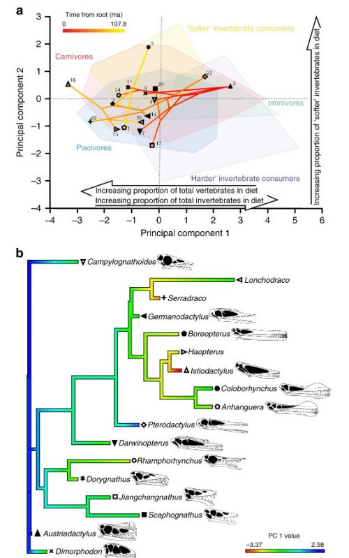
Sometimes it goes a little too far, though — for example, this illustration is way too dense to be useful, but it it interesting.

That is not a spider

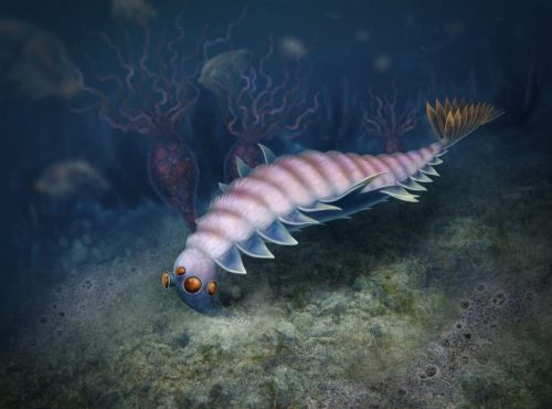
Grrr. The CBC got me excited with a headline about “the granddaddy of spiders”. It’s not a spider. It’s a Cambrian chelicerate, which ought to be cool news enough without pretending it’s some kind of familiar organism. At least it wasn’t SciTech, which called it a frightening 500-million year old predator” or LiveScience, which called it a “nightmare creature”. C’mon, people. It was a couple of centimeters long. I do not like this pop sci nonsense that has to jack up the significance of a discovery by pretending it was scary. Does this look scary to you?

a–c, Reconstructions. a, Lateral view. b, Dorsal view (the gut has been removed for clarity). c, Isolated trunk exopod. an, anus; lam, lamellae.

At least the article by the discoverers is sensible. This is an early Cambrian chelicerate with those big old feeding appendages at the front of the head (which spiders also have) and with modified limb appendages that resemble book lungs (also a spider trait), but they are most definitely not spiders. They are their own beautiful clade, and cousins of Mollisonia plenovenatrix might have been spider ancestors, but calling them spiders is like excavating an ancient fish and calling it a mammal. Very misleading.

Yes, I’m being pedantic. It matters. Let’s not diminish the diverse chelicerates by calling them spider wanna-bes.

Here’s the abstract for the paper.

The chelicerates are a ubiquitous and speciose group of animals that has a considerable ecological effect on modern terrestrial ecosystems—notably as predators of insects and also, for instance, as decomposers. The fossil record shows that chelicerates diversified early in the marine ecosystems of the Palaeozoic era, by at least the Ordovician period. However, the timing of chelicerate origins and the type of body plan that characterized the earliest members of this group have remained controversial. Although megacheirans have previously been interpreted as chelicerate-like, and habeliidans (including Sanctacaris) have been suggested to belong to their immediate stem lineage, evidence for the specialized feeding appendages (chelicerae) that are diagnostic of the chelicerates has been lacking. Here we use exceptionally well-preserved and abundant fossil material from the middle Cambrian Burgess Shale (Marble Canyon, British Columbia, Canada) to show that Mollisonia plenovenatrix sp. nov. possessed robust but short chelicerae that were placed very anteriorly, between the eyes. This suggests that chelicerae evolved a specialized feeding function early on, possibly as a modification of short antennules. The head also encompasses a pair of large compound eyes, followed by three pairs of long, uniramous walking legs and three pairs of stout, gnathobasic masticatory appendages; this configuration links habeliidans with euchelicerates (‘true’ chelicerates, excluding the sea spiders). The trunk ends in a four-segmented pygidium and bears eleven pairs of identical limbs, each of which is composed of three broad lamellate exopod flaps, and endopods are either reduced or absent. These overlapping exopod flaps resemble euchelicerate book gills, although they lack the diagnostic operculum. In addition, the eyes of M. plenovenatrix were innervated by three optic neuropils, which strengthens the view that a complex malacostracan-like visual system might have been plesiomorphic for all crown euarthropods. These fossils thus show that chelicerates arose alongside mandibulates as benthic micropredators, at the heart of the Cambrian explosion.

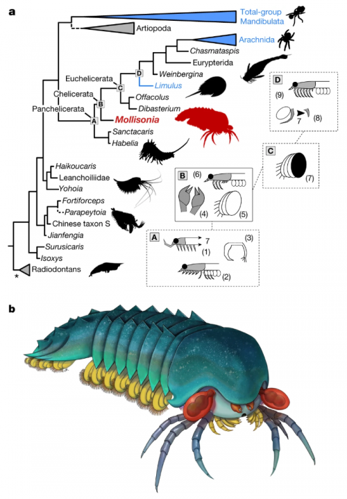
I think this diagram illustrates the relationship of M. plenoventrix to spiders well.

a, Simplified consensus tree of a Bayesian analysis of panarthropod relationships. This tree is based on a matrix of 100 taxa and 267 characters. Extant taxa are in blue; dashed branches represent questionable groupings. Asterisk shows that the radiodontans resolved as paraphyletic. This analysis excludes pycnogonids, but this had little effect on the topology. The letters A to D at the basal panchelicerate nodes refer to boxes on the right, and summarize the appearances of major morpho-anatomical features: (1) extension of cephalic shield, including a seventh tergite; (2) cephalic limbs all co-opted for raptorial and masticatory functions, and reduction of some trunk endopods; (3) dissociation of the exopod from the main limb branch; (4) presence of chelicerae; (5) trunk exopods made of several overlapping lobes; (6) some cephalic limbs differentiated as uniramous walking legs; (7) multi-lobate exopod covered by sclerite (operculum); (8) reduction of seventh cephalic appendage pair; and (9) all post-frontal cephalic limb pairs are uniramous walking legs. b, Life reconstruction. Drawing by J. Liang, copyright Royal Ontario Museum

Not a spider, but still cute and adorable.


Aria C, Caron J-B (2019) A middle Cambrian arthropod with chelicerae and proto-book gills. Nature https://doi.org/10.1038/s41586-019-1525-4.

I don’t understand it, therefore nobody does

We’re going to see a wave of ignorance prompted by David Gelernter’s profession of foolishness, aren’t we? Every fool in the world who hears that guy’s nonsense is now inspired to spew out some nonsense of their own.

One example is Barbara Kay, who I’ve never heard of before, pontificating in the National Post that “there’s one mystery we still can’t explain”. Only one? I can think of lots. But the fact that there are still questions in the world does not mean that all the answers we have are wrong.

Her point is especially bad, because she singles out one thing that she thinks is false, and she is wrong about it.

The human brain and the power of speech put humans way beyond the boundaries of Darwin’s own three critical criteria for natural selection, which; i) may expand an animal’s power only to a point where it has survival advantage — and no further; ii) cannot produce changes that are “injurious” to the animal; and iii) cannot produce a “specially developed organ” that is useless to an animal at the time it develops. If a Neanderthal brain three times the size of any primate’s and a unique capacity for speech do not constitute “specially developed organs,” what does?

OK. Start with Darwin: he’s not our infallible prophet. He got a lot wrong, and remember, he was writing 150 years ago. You can demonstrate Darwin’s errors all you want, and modern scientists will just shrug and say, “So?”

Kay’s second error, though, is that she overlooked the meaning of her subject, natural selection. Evolution is not synonymous with natural selection, and showing that something could not have evolved by natural selection does not refute the idea that it evolved by some other mechanism. Even if we take those three points as given, it does not negate the idea of evolution.

Third error: she has not demonstrated that point (i) means natural selection could not have occurred. Where does the survival advantage of speech stop? It seems to me that the initiation of speech with grunts and crude vocalizations could only be improved, and improved continuously, by natural selection. Speech that enabled better hunting could lead to speech that is used for love poetry, or describing geography, or telling scary stories around the campfire, or expressing philosophical thoughts. She has not demonstrated any barrier which would impede the action of natural selection.

Fourth error: The brain isn’t that special (ii). All animals have one (well, we could call sponges and jellyfish exceptions). Our ancestors had one that could visualize the environment and the future, allow for sophisticated socialization, and permitted all kinds of communication shy of speech. Speech capability builds on structures that are already present in a multitude of animals.

Fifth error: brains that could process information in a complex way before speech evolved were not useless to our ancestors (iii), even if they couldn’t speak.

Sixth and biggest, most common error in creationists: the failure of their imaginations and ignorance of the evidence does not support their claim that the science is wrong. I can’t imagine how Barbara Kay manages to type words on a machine, but I think it’s clear that she did. Probably. I can’t rule out the possibility that an editor filtered the output of a monkey pounding on a keyboard, but it’s more likely that her essay was produced by a human being who simply knows nothing about biology.

Actually, I fail to see a single thing in this paper that would require any textbook rewriting at all

Something about this title, “Evolutionary discovery to rewrite textbooks”, put me on edge. It’s a common trope to announce that your specific discovery is going to revolutionize everything, therefore you deserve more attention and glory and grant money. (Creationists seem not to understand this dynamic, though — they think scientists stick with the safe, and they think wrong, theory for all the money, when everyone knows a good, robust insight that changes minds is where the glory lies).

It’s in phys.org, though, which is generally a garbage fire of badly butchered summaries of papers, so that fact alone primes me to think the summary writer didn’t really understand the paper.

But then, it quotes the study author.

“This technology has been used only for the last few years, but it’s helped us finally address an age-old question, discovering something completely contrary to what anyone had ever proposed.”

“We’re taking a core theory of evolutionary biology and turning it on its head,” she said.

“Now we have an opportunity to re-imagine the steps that gave rise to the first animals, the underlying rules that turned single cells into multicellular animal life.”

Professor Degnan said he hoped the revelation would help us understand our own condition and our understanding of our own stem cells and cancer.

Aaaargh, no. They’re claiming that multicellular animals did not evolve from a single-celled ancestor resembling a choanoflagellate, a small protist with a single, prominent flagellar “propellor”, which many scientists consider to resemble one of the cells of sponges, called choanocytes. The idea is that the first multicellular animal would have arisen from colonies of choanoflagellates ganged together, with specialization of other cell types evolving later.

It’s fine and interesting that these investigators are proposing an alternative model, but this is not a “core theory of evolutionary biology”. It’s a likely hypothesis for the origin of a lineage of organisms we selfishly consider important, because it includes us, but it doesn’t actually revolutionize any part of the theory of evolution. Way too many scientists fail to grasp that there are the theories of evolution, that explain general mechanisms of the process, and that there are a multitude of facts of evolution, which are the instances in history that led to the specific distribution of evolutionary outcomes.

It’s like how there are physics theories that explain general phenomena: your car operates on all kinds of rules about force and acceleration and combustion. If you one day discover a shortcut on your commute to work, it may be an important personal discovery, but you don’t get to crow triumphantly about turning a core theory of automotive engineering and mechanics on its head.

But then, this is phys.org…I guess I’d better read the original paper.

It really is an interesting paper. It looks at the transcriptome of different cell types in sponges and compares them to the transcriptome of a choanoflagellate, and is basically addressing the reliability of a conclusion drawn from an observed phenotype, versus a conclusion drawn from patterns of gene expression. It’s going to argue that gene expression ought to be more fundamental, and I can sort of agree (while also seeing some problems of interpretation), but it then leaps to evolutionary conclusions from cell types, which I don’t find persuasive at all.

To summarize briefly: there are roughly three cell types in a sponge: 1) the choanocytes, which are flagellar motors that drive water flow through the animal; 2) pinacocytes, which form epithelial sheets that line the outside and insides; and 3) archaeocytes, which are found in a gooey mesenchymal smear between the layers of pinacocytes. A sponge is kind of sandwich, where the pinacocytes form the bread, the archaeocytes are the jelly, and the choanocytes are…damn, my analogy is breaking down. The choanocytes are imbedded in the bread and stir the surroundings.

The question then is, what genes are being expressed in these three cell types? And the answer is, well, lots of genes, and many of them are being differentially expressed — that is, there are genes unique to each cell type that reflect their general role.

We find that archaeocytes significantly upregulate genes involved in the control of cell proliferation, transcription and translation, consistent with their function as pluripotent stem cells. By contrast, choanocyte and pinacocyte transcriptomes are enriched for suites of genes that are involved in cell adhesion, signalling and polarity, consistent with their role as epithelial cells.

Now though, they raise an evolutionary question. They identify all these genes, and then ask which are common to single celled eukaryotes, which are common to multicellular animals, and which are unique to sponges. This gives a rough estimate of the evolutionary age of these genes; the first category is the oldest, found in pre-metazoan organisms, the second category may have arisen at the approximate time of origin of the metazoan lineage, and the third category would have appeared later as the sponge lineage specialized. They determined the relative contributions of these three categories to the patterns of gene expression in the three cell types.

The A. queenslandica [the sponge species] genome comprises 28% pre-metazoan, 26% metazoan and 46% sponge-specific protein-coding genes. We find that 43% of genes significantly upregulated in choanocytes are sponge-specific, which is similar to the proportion of the entire genome that is sponge-specific. By contrast, 62% of genes significantly upregulated in the pluripotent archaeocytes belong to the evolutionarily oldest pre-metazoan category, significantly higher than the 28% of genes for the entire genome. As with archaeocytes, pinacocytes express significantly more pre-metazoan and fewer sponge-specific genes than would be expected from the whole-genome profile.

Does this diagram help interpret that?

a, Phylostratigraphic estimate of the evolutionary age of coding genes in the A. queenslandica genome. b–d, Estimate of gene age of differentially expressed genes in choanocytes (b, top), archaeocytes (c, top) and pinacocytes (d, top) and the enrichment of phylostrata relative to the whole genome (b–d, bottom). Asterisks indicate significant difference (two-sided Fisher’s exact test P <0.001) from the whole genome. The enrichment values (log-odds ratio) for: choanocytes (b; n = 10) are sponge-specific (−0.0089, P = 0.7747), metazoan (–0.0361, P = 0.9958) and premetazoan (0.0439, P = 0.0004) genes; archaeocytes (c; n = 15) are sponge-specific (−0.5634, P = 1.33 × 10−133), metazoan (−0.1923, P = 1.04 × 10⁻¹⁸) and premetazoan (0.6772, P = 0); and pinacocytes (d; n = 6) are sponge-specific (−0.2173, P = 5.23 × 10⁻¹³), metazoan (−0.0008, P = 0.5231) and premetazoan (0.2359, P = 3.07 × 10−36).

OK, maybe not. Shorter summary: archaeocytes express lots of old genes, which makes sense, given we already had it explained that they’re enriched for genes involved in control of cell proliferation, transcription and translation, which are also ancient, primitive functions. Pinacocytes are also doing fairly basic things, and like the archaeocytes, are switching on basic essential genes. The choanocytes, though, are exceptional, and are turning on lots of sponge-specific genes that evolved after sponges diverged from other metazoans. The conclusion: choanocytes are switching on derived spongey genes that evolved for a spongey lifestyle, while archaeocytes are more generic, switching on a universal toolkit for adaptable stem cells, which have to be able to change their roles to become any of the three cell types.

They also make the point that the choanoflagellate gene expression pattern is most similar to that of the archaeocytes. From these observations, they argue that the ancestral metazoan more resembled a stem cell, like an archaeocyte, than a choanoflagellate.

…we posit that the ancestral metazoan cell type had the capacity to exist in and transition between multiple cell states in a manner similar to modern transdifferentiating and stem cells. Recent analyses of unicellular holozoan genomes support this proposition, with some of the genomic foundations of pluripotency being established deep in a unicellular past. Genomic innovations unique to metazoans—including the origin and expansion of key signalling pathway and transcription factor families, and regulatory DNA and RNA classes—may have conferred the ability of this ancestral pluripotent cell to evolve a regulatory system whereby it could co-exist in multiple states of differentiation, giving rise to the first multicellular animal.

And that’s where they lose me. I don’t buy their interpretation.

First, organisms do not evolve by descent from cell types. The whole genome is passed down. That choanocytes express a certain subset of genes is irrelevant — they carry the whole suite of genes, as do the pinacocytes and archaeocytes. You wouldn’t do a gene expression profile of human brain cells and compare it to the profile for mesenchymal cells, and then argue that brain cells are weird and unique, therefore we didn’t evolve from brain cells. Of course we didn’t! Your assumptions make no sense.

Second, what they found was that choanoflagellates exhibit a broader, more general pattern of gene expression than sponge choanocytes. This makes sense. Sponges are truly multicellular, with specialized subsets of cells that carry out specific roles, while choanoflagellates are unicellular, where each cell has to be a jack-of-all-trades. Yes, a choanoflagellate is going to have to use all of its genes, while individual cells in the sponge have the luxury of focusing on a smaller set of jobs. Choanocytes are specialized, they’re going to have a different expression profile than a generalist cell.

Third, choanoflagellates have an expression profile similar to an archaeocyte…but this is not in contradiction to their phenotype of having a flagellar collar. The metazoan ancestor could still have been a choanocyte, and nothing in this evidence contradicts that possibility!

All they’re really saying when you get right down to it is that metazoan ancestor had to have the capacity to generate diverse cell types later in its evolution, which is kind of obvious. They’re also arguing that the ancestral metazoan could not have been as locked in and limited in its repertoire of functions as a choanocyte in an extant sponge, which, again, is a given. What is not obvious is that the ancestor had to have been archaeocyte-like in form and function.

I also get the impression that the authors have been soaking in the transcriptomics literature and practice to the extent that they’ve lost sight of the bigger picture. They’d only have an argument if descendant forms were derived from the RNA of their ancestors…which actually would revolutionize the theory of evolution! Fortunately, they are not making that claim at all.


Sogabe S, Hatleberg WL, Kocot KM, Say TE, Stoupin D, Roper KE, Fernandez-Valverde SL, Degnan SM & Degnan BM (2019) Pluripotency and the origin of animal multicellularity. Nature https://doi.org/10.1038/s41586-019-1290-4

Teams of Memes, bursting from the seams

Image courtesy of the googles.

Daniel Dennett’s From Bacteria to Bach and Back is a lengthy and winding journey. It is characterized (including by its publisher) as a general explanation of the evolution of minds and various peculiar mental functions, consciousness and language being the two most hotly discussed by philosophers, but there’s a better way to read it. As its best, the book is a tour of Dennett’s personal philosophical repertoire, illustrating how ideas from his books and papers fit together.

Dennett’s general theory of the development of genetics stems from his broad theory of memes, where a meme is any informational entity that can be transmitted and replicated. The rough idea is that minds are meme-machines in the way that organisms are gene-machines (in Dawkins’ analogy of the gene’s-eye-view). This is a fruitful analogy, in some respects, though I think it can and should draw some skepticism from readers. I’ll return to those worries later.

The basic building blocks of Dennett’s view are indicated by gestures and short explanations, which is a challenge since he’s spent so much time discussing and arguing for them elsewhere in his work. In any case, there are really two that it is important to understand.

[Read more…]

Protists, not animals

I’ve written about the spectacular phospatized embryos of the Doushantuo formation before. It’s a collection of exceptionally well preserved small multicellular organisms, so well preserved that we can even look at cellular organelles. And they’re pre-Cambrian, as much as 630 million years old.

They’ve been interpreted as fossilized embryos for which we have no known adult forms. They certainly look like embryos, but one thing has always bothered me — they all look like blastula-stage embryos at various points in their early divisions, and the absence of later stages was peculiar: how did gastrulae and neurulae and other stages avoid getting preserved?

One explanation was that we weren’t seeing metazoan fossils at all — they were colonies of large bacteria. That’s disappointing if you have an animal bias, but still cool — as I pointed out then, it just highlights the fact that the transition from single-celled to multi-celled life isn’t that remarkable.

Now we have another alternative explanation that seems even better to me: they aren’t animals, and they aren’t bacteria, they’re protists. Some of the Doushantuo specimens are rather peanut-shaped, and others are vermiform, odd for an animal embryo, but entirely compatible with the idea that these are encysted stages of propagating protists.

Here are some of these oddly shaped Doushantuo specimens.

i-9bc3e359c5dbbed4c0f7e4e077a77157-tianzhushania.jpeg
Tianzhushania from the Ediacaran Doushantuo Formation, Datang Quarry, Weng’an, Guizhou Province, China. (A) Regular and (B to J) irregular forms, the latter interpreted to be in the germinating stage: MESIG 10022 [(A) SEM micrograph]; MESIG 10023 [(B) SEM micrograph (19)]; MESIG 10024 [(C) SEM micrograph (19)]; MESIG 10021 [(D) SEM micrograph]; SMNH X 4447 [(E) to (G) srXTM renderings]; SMNH X 4448 [(H) to (J) srXTM renderings]. (A) Surface of regular globular specimen shows envelope structure, to be compared with the similar envelope structure in (B) to (D). [(B) and (C)] Germinating specimens show protruding tubes and envelope structure. (D) Peanut-shaped specimen shows envelope structure. (E) Isosurface rendering of peanut-shaped specimen. (F) Orthoslice through (E). (G) Detail of approximate level in (F), showing cellular units. (H) Isosurface rendering of peanut-shaped specimen. (I) Orthoslice through (H). (J) Detail of approximate level in (I), showing cellular units. There is a progressive individuality of cellular units toward the periphery, including detachment of single- and oligocellular units (arrows).
i-c2ed0d62b380987fc5477a57e4735e06-lifecycle.jpeg
Proposed life cycle of Tianzhushania through hypertrophic growth of mother cell, encystment in multilayered wall, palintomic cleavage resulting in a tightly packed mass of pre-propagules, germination by opening of outer cyst wall, and release of prop- agules by degradation of inner cyst wall. Shown is the role of the outer and inner cyst walls in forming the peanut-shaped germination stages (see also modern mesomycetozoean examples in fig. S7). The outer cyst wall (seldom preserved) is indicated in black; the inner cyst wall dark is indicated in gray.

Their proposed explanation convinces me. These were protists that were single-celled in their free-living stage which would periodically grow hypertrophically and encyst, forming a capsule containing the dividing cells. These cells would replicate at differnt rates, forming zones of maturation; eventually, the cyst would rupture, released a cloud of propagules, or spores, and the life cycle would begin again.

That would explain a lot about the distribution of forms in these phosphatized specimens — we don’t find any gastrulating embryos because there never were any. These weren’t animals, period!

They belong outside crown-group Metazoa, within total-group Holozoa (the sister clade to Fungi that includes Metazoa, Choanoflagellata, and Mesomycetozoea) or perhaps on even more distant branches in the eukaryote tree. They represent an evolutionary grade in which palintomic cleavage served the function of producing propagules for dispersion.

That’s still very interesting, and again, it reminds us that the transition to multicellularity had many antecedents and could have been reached by many different paths.


Huldtgren T, Cunningham JA, Yin C, Stampanoni M, Marone F, Donoghue PC, Bengtson S (2011) Fossilized nuclei and germination structures identify Ediacaran “animal embryos” as encysting protists. Science 334(6063):1696-9.

(Also on FtB)

Why do women menstruate?

Menstruation is a peculiar phenomenon that women go through on a roughly monthly cycle, and it’s not immediately obvious from an evolutionary standpoint why they do it. It’s wasteful — they are throwing away a substantial amount of blood and tissue. It seems hazardous; ancestrally, in a world full of predators and disease, leaving a blood trail or filling a delicate orifice with dying tissue seems like a bad idea. And as many women can tell you, it’s uncomfortable, awkward, and sometimes debilitating. So why, evolution, why?

One assumption some people might make is that that is just the way mammalian reproduction works. This isn’t true! Most mammals do not menstruate — they do not cycle their uterine linings, but instead only build up a thickened endometrium if fertilization occurs, which looks much more efficient. Of the mammals, only most primates, a few bats, and elephant shrews are among the lucky animals that menstruate, and as you can see from the phylogeny, the scattered diversity of menstruating mammals implies that the trait was not present ancestrally — we primates acquired it relatively late.

i-58b845535df701c0ab264735f8049106-menstrual_phylogeny-thumb-500x494-71491.jpeg

Phylogeny showing the distribution of menstruation in placental mammals and the inferred states of ancestral lineages. Menstruating species/lineages are colored in pink, non- menstruating species/lineages in black. Species in which the character state is not known are not colored, and lineages of equivocal state are represented with black lines. Monodelphis represents the outgroup. Inference of ancestral states was performed in MacClade 4 by the parsimony method. Note that there is strong evidence for three independent originations of menstruation among placental mammals.

I suppose we could blame The Curse on The Fall, but then this phylogeny would suggest that Adam and Eve were part of a population of squirrel-like proto-primates living in the early Paleocene. That’s rather unbiblical, though, and what did the bats and elephant shrews do to deserve this?

There are many explanations floating around. One is that it’s a way to flush out nasty pathogens injected into the reproductive tract by ejaculating males — but that phenomenon is ubiquitous, so you have to wonder why only a few species bother. Another explanation is that it’s more efficient to get rid of the endometrium when not using it, than to maintain it indefinitely; but this is a false distinction, because other mammals don’t maintain the endometrium, they just build it up in response to fertilization. And finally, another reason is that humans have rather agressive embryos that implant deeply and intimately with the mother’s tissues, and menstruation “preconditions” the uterine lining to cope with the stress. There is, unfortunately, no evidence that menstruation provides any boost to the ‘toughness’ of the uterus at all.

A new paper by Emera, Romero, and Wagner suggests an interesting new idea. They turn the question around: menstruation isn’t the phenomenon to be explained, decidualization, the production of a thickened endometrial lining, is the key process.

All mammals prepare a specialized membrane for embryo implantation, the difference is that most mammals exhibit triggered decidualization, where the fertilized embryo itself instigates the thickening, while most primates have spontaneous decidualization (SD), which occurs even in the absence of a fertilized embryo. You can, for instance, induce menstruation in mice. By scratching the mouse endometrium, they will go through a pseudopregnancy and build up a thickened endometrial lining that will be shed when progesterone levels drop. So the reason mice don’t menstruate isn’t that they lack a mechanism for shedding the endometrial lining…it’s that they don’t build it up in the first place unless they’re actually going to use it.

So the question is, why do humans have spontaneous decidualization?

The answer that Emera suggests is entirely evolutionary, and involves maternal-fetal conflict. The mother and fetus have an adversarial relationship: mom’s best interest is to survive pregnancy to bear children again, and so her body tries to conserve resources for the long haul. The fetus, on the other hand, benefits from wresting as much from mom as it can, sometimes to the mother’s detriment. The fetus, for instance, manipulates the mother’s hormones to weaken the insulin response, so less sugar is taken up by mom’s cells, making more available for the fetus.

Within the mammals, there is variation in how deeply the fetus sinks its placental teeth into the uterus. Some species are epithelochorial; the connection is entirely superficial. Others are endotheliochorial, in which the placenta pierces the uterine epithelium. And others, the most invasive, are hemochorial, and actually breach maternal blood vessels. Humans are hemochorial. All of the mammalian species that menstruate are also hemochorial.

That’s a hint. Menstruation is a consequence of self-defense. Females build up that thickened uterine lining to protect and insulate themselves from the greedy embryo and its selfish placenta. In species with especially invasive embryos, it’s too late to wait for the moment of implantation — instead, they build up the wall pre-emptively, before and in case of fertilization. Then, if fertilization doesn’t occur, the universal process of responding to declining progesterone levels by sloughing off the lining occurs.

Bonus! Another process that goes on is that the lining of the uterus is also a sensor for fetal quality, detecting chromosomal abnormalities and allowing them to be spontaneously aborted early. There is some evidence for this: women vary in their degree of decidualization, and women with reduced decidualization have been found to become pregnant more often, but also exhibit pregnancy failure more often. So having a prepared uterus not only helps to fend off overly-aggressive fetuses, it allows mom a greater ability to be selective in which fetuses she carries to term.

The authors also have a proposed mechanism for how menstruation could have evolved, and it involves genetic assimilation. Genetic assimilation is a process which begins with an environmentally induced phenotype (in this case, decidualization in response to implantation), which is then strengthened by genetic mutations that stabilize the phenotype — phenotype first, followed by selection for the mutations that reinforce the phenotype. They make predictions from this hypothesis. In species that don’t undergo SD, embryo implantation triggers an elevation of cyclic AMP in the endometrium that causes growth of the lining. If genetic assimilation occurred, they predict that what happened in species with SD was the novel coupling of hormonal signaling to the extant activation process.

If either of these models were correct, we would expect an upregulation of cAMP- stimulating agents in response to pro- gesterone in menstruating species like humans, but not in non-menstruating species such as the mouse.

Results from experiments like those described above will elucidate the evolutionary pathway from induced to spontaneous decidualization, allowing us to answer long-unanswered questions about the evolutionary significance of menstruation. In addition, they will provide mechanistic insights that might be useful in the treatment of common reproductive disorders such as endometriosis, endometrial cancer, preeclampsia, and recurrent pregnancy loss. These disorders involve dysfunctional endometrial responses during the menstrual cycle and pregnancy. Thus, clarifying mechanisms of the normal endometrial response to maternal hormones, i.e. SD, will facilitate identification of genes with abnormal function in women with these disorders. An analysis of how SD came about in evolution can aid in identifying these critical molecular mechanisms.

Evolution, genetic assimilation, a prediction from an evolutionary hypothesis, and significant biomedical applications … that all sounds powerful to me.


Emera D, Romero R, Wagner G (2011) The evolution of menstruation: A new model for genetic assimilation: Explaining molecular origins of maternal responses to fetal invasiveness. Bioessays 34(1):26-35.

(Also on FtB)

Creationist abuse of cuttlefish chitin

A few weeks ago, PLoS One published a paper on the observation of preserved chitin in 34 million year old cuttlebones. Now the Institute for Creation Research has twisted the science to support their belief that the earth is less than ten thousand years old. It was all so predictable. It’s a game they play, the same game they played with the soft tissue preserved in T. rex bones. Here’s how it works.

Compare the two approaches, science vs. creationism. The creationists basically insert one falsehood, generate a ludicrous conflict, and choose the dumbest of the two alternatives.

The Scientific Approach

find traces of organic material in ancient fossils

Cool! We have evidence of ancient biochemistry!

Science!

The Creationist Approach

declare it impossible for organic material to be ancient

steal other people’s discovery of organic material in ancient fossils

Cool! Declare that organic material must not be ancient, because of step 1, which we invented

Throw out geology, chemistry, and physics because they say the material is old

Profit! Souls for my Lord Arioch!

You see, the scientists are aware of the fact that organic materials degrade over time, but recognize that we don’t always know the rate of decay under all possible conditions. When we find stuff that hasn’t rotted away or been fully replaced by minerals, we’re happy because we’ve got new information about ancient organisms, and we may also be able to figure out what mechanisms promoted the preservation of the material.

The creationists start with dogma — in this case, a false statement. They declare

Chitin is a biological material found in the cuttlebones, or internal shells, of cuttlefish. It has a maximum shelf life of thousands of years…

and

Because of observed bacterial and biochemical degradation rates, researchers shouldn’t expect to find any original chitin (or any other biomolecule) in a sample that is dozens of millions of years old–and it therefore should be utterly absent from samples deposited hundreds of millions of years ago. Thus, the chitin found in these fossils refutes their millions-of-years evolutionary interpretation, just as other fossil biomolecules already have done.

But wait. How do they know that? The paper they are citing says nothing of the kind; to the contrary, it argues that while rare, other examples of preserved chitin have been described.

Detection of chitin in fossils is not frequent. There are reports of fossil chitin in pogonophora, and in insect wings from amber. Chitin has also been reported from beetles preserved in an Oligocene lacustrine deposit of Enspel, Germany and chitin-protein signatures have been found in cuticles of Pennsylvanian scorpions and Silurian eurypterids.

So the paper is actually saying that the “maximum shelf life” of chitin is several tens of millions of years. And then they go on to describe…chitin found in Oligocene cuttlefish, several tens of millions of years old. The creationists are busily setting up an imaginary conflict in the evidence, a conflict that does not exist and is fully addressed in the paper.

The creationists do try to back up their claims, inappropriately. They cite a couple of papers on crustacean taphonomy where dead lobsters were sealed up in anoxic, water- and mud-filled jars; they decayed. Then they announce that there’s only one way for these cuttlebones to be preserved, and that was by complete mineralization, and the cuttlebones in the PLoS One paper were not mineralized.

…mineralization–where tissues are replaced by minerals–is required for tissue impressions to last millions of years. And the PLoS ONE researchers verified that their cuttlebone chitin was not mineralized.

Funny, that. You can read the paper yourself. I counted 14 uses of the words “mineralized” and “demineralized”. They state over and over that they had to specifically demineralize the specimens in hydrochloric acid to expose the imbedded chitin. And of course the chitin itself hadn’t been mineralized, or it wouldn’t be chitin anymore! Did the creationists lie, or did they just not understand the paper?

The scientists also do not claim that the chitin has not been degraded over time. They actually document some specific, general properties of decay in the specimens.

β-chitin is characterized by parallel chains of chitin molecules held together with inter-chain hydrogen bonding. The OH stretching absorbance, at about 3445 cm−1 in extant chitin, is diminished in the fossil and shifted to lower wavenumbers, showing that the specimen is losing OH by an as yet undetermined mechanism. The N-H asymmetric stretching vibration is shifted to slightly lower wavenumbers, showing that it is no longer hydrogen bonding exactly as in extant specimens. Changes in the region 2800-3600 cm−1 indicate that biomolecules have been degraded via disruption of interchain hydrogen bonds.

So, yes, the creationists seem to have rather misrepresented what the paper said. Here’s another blatant example of lying about the contents of the paper.

The question they did not answer, however, is why the original organic chitin had not completely fallen apart, which it would have if the fossils with it were 34 million years old…

Actually, they did. A substantial chunk of the discussion was specifically about that question, a consideration of the factors that contributed to the preservation. It was a combination of an anoxic environment, the presence of molecules that interfered with the enzymes that break down chitin, and the structure of cuttlebone, which interleaves layers containing chitin with layers containing pre-mineralized aragonite.

In vivo inorganic-organic structure of the cuttlebone, in combination with physical and geochemical conditions within the depositional environment and favorable taphonomic factors likely contributed to preservation of organics in M. mississippiensis. Available clays within the Yazoo Clay in conjunction with suboxic depositional environment may have facilitated preservation of original organics by forming a physical and geochemical barrier to degradation. One key to the preservation of organic tissues, in particular chitin and chitosan, is cessation of bacterial degradation within environments of deposition. Bacterial breakdown of polymeric molecules is accomplished through activities of both free extracellular enzymes (those in the water column) and ektoenzymes (those on the surface of the microbial cell) such as chitinases. Chitinases function either by cleaving glycosidic bonds that bind repeating N-acetyl-D-glucosamine units within chitin molecules or by cleaving terminal N-acetyl-D-glucosamine groups. These enzymes adsorb to the surface of clay particles, which inactivates them. Strong ions in solution like iron may act in the same manner. Once bound to functional groups within these polymeric molecules, Fe2+ ions prevent specific bond configuration on the active-site cleft of specific bacterial chitinases and prevents hydrolysis, thus contributing to preservation.

Organic layers within cuttlebones are protected by mineralized layers, similar to collagen in bones, and this mineral-organic interaction may also have played a role in their preservation. Specimens of M. mississipiensis show preserved original aragonite as well as apparent original organics. These organics appear to be endogenous and not a function of exogenous fungal or microbial activity. Fungi contain the γ form of chitin not the β allomorph found in our samples. Also, SEM analyses shows there is no evidence of tunneling, microbes, or wide-spread recrystallization of the aragonite. Therefore the chitin-like molecules detected in fossil sample are most likely endogenous. Similar to collagen in bone, perhaps, organics could not be attacked by enzymes or other molecules until some inorganic matrix had been removed.

Like I always say, never ever trust a creationists’ interpretation of a science paper: they don’t understand it, and they are always filtering it through a distorting lens of biblical nonsense. They make such egregious errors of understanding that you’re always left wondering whether they are actually that stupid, or that sleazily dishonest. Or both.

But imagine if the creationists hadn’t screwed up royally in reading the paper, if they had actually found an instance of scientists being genuinely baffled by a discovery that should not be. What if there was actually good reason to believe that chitin could not last more than ten thousand years?

Then the only sensible interpretation of this observation of 34 million year old chitin would be that the prior estimation of the shelf-life of chitin was wrong, and that it could actually last tens of millions of years. What the creationists want to do is claim that that minor hypothetical is actually correct, and that instead the entirety of nuclear physics, geology, radiochemistry, and modern cosmology is wrong. On the one hand, uncertain details about the decay of one organic molecule; in the other, entire vast fields of science, already verified, and with complex modern technologies built on their operation…and which hand would the creationists reject? The trivial one, of course.

I’m leaning towards “stupid” as the explanation for their bad arguments.


Oh, after I started this dissection, I discovered someone had already beaten me to it: here’s another analysis of the creationist misinterpretations.

(Also on FtB)

Gals and show mares

This video has been going around — it’s a group of women talking about the importance of evolution to the biological sciences.

I confess to cringing in a few places — there’s too much ready equation of evolution with natural selection — but I certainly wouldn’t question the competence of these accomplished scientists, even if I might argue with them a bit.

But now the clowns at Uncommon Descent have discovered it and given their assessment.

It shows sixteen female academics or science writers, mostly young, whose enthusiasm for evolution is so overwrought that they turn themselves into propagandists.

Eager to show how well they have been trained, they are like show mares who trot around the paddock jumping over each gate in turn. All the while they give the camera a look that says: “Aren’t I good?”

And then the conclusion:

Here, we’d wondered who would be the next Lynn Margulis. Our scouts can now save time by crossing these gals off.

“Gals”? Really? And since when do creationist hacks get to cross “gals” off the rolls of worthy scientists?

That’s right there in the article. There is worse in the comments; I know the site isn’t entirely responsible for what commenters say, but this is from one widely known freakish creationist who agrees with the sentiment in the article, that these women won’t cut it as real scientists. (There are also others that disagree with this guy; no one seems to have noted the patronizing attitude of the article itself.)

There is however a liberal establishment with a agenda to promote women and this means over more deserving men. Affirmative action , openly/secret, is powerful in nOrth america.
They want women to be as smart as men in these perceived smarter things.
They think it should be at least 50/50.
However it ain’t and it never will.

(Also on FtB)

Anti-caturday post

There is actually a cat in this video. Notice, though, that it only appears briefly in the beginning, looks bored, and apathetically wanders off screen. Why? Because the rest of the video features something far more exciting and bizarre than a mere cat: it’s all about zombie fish, their brains infected with trematode parasites. The cat knows that it cannot compete, unless it goes off and gets its brain tainted with some freaky strange parasite to give it some character.

Another interesting thing about it is that this video is an attempt to get funding for science research. If you feel like promoting more research into how to infect brains and make zombies, donate!

(Also on FtB)