BIRDIE!

Time to dig up another fossil. It’s a bird, but it has no connection to the sabre-toothed kitty cat I posted yesterday — this is a 80 million year old bird, Navaornis hestiae, written up in a Nature article, Cretaceous bird from Brazil informs the evolution of the avian skull and brain. It looks like a real bird to me.

a,b, Photograph (a) and interpretive drawing (b) of the exposed side of the holotype of N. hestiae (MPM-200-1) in left lateral view. c, Micro-computed tomography rendering of MPM-200-1 in right ventral–lateral view. Scale bar, 10 mm.

It has a fairly big brain, with some differences in structure from modern birds — it has a smaller motor control area, so while it had the capacity for complex behavior, it may not have been as agile in the air as birds today. It’s intermediate in brain complexity between Archaeopteryx and extant birds.

a, Three-dimensional reconstruction of the endocranial morphology of N. hestiae from MPM-200-1 and MPM-334-1. Portions deriving from MPM-200-1 and MPM-334-1, as well as the reconstruction process, are explained in the Methods and Extended Data Fig. 7. b, Evolution of endocranial morphology across Pennaraptora. Numbers in the coloured boxes refer to the degree of expansion of each of the main neuroanatomical and sensorial regions for each taxon. Brown arrows in b depict the orientation of the foramen magnum.

Cambridge invested a bit in publicizing this discovery, with a nice fancy video.

KITTY!

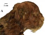
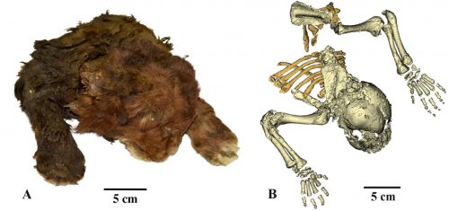
External appearance of three-week-old heads of large felid cubs, right lateral view: (A) Homotherium latidens (Owen, 1846), specimen DMF AS RS, no. Met-20-1, frozen mummy, Russia, Republic of Sakha (Yakutia), Indigirka River basin, Badyarikha River; Upper Pleistocene

I understand that the internet likes cats, so here’s one, a 30,000 year old mummified sabre-toothed kitten.

It has a distinctively large mouth and massive neck muscles, but the canine teeth haven’t elongated yet — they say the age is equivalent to a 3 week old lion cub. I would guess that sabre-tooth canines might interfere with nursing.

The frozen mummy of Homotherium latidens (Owen, 1846), specimen DMF AS RS, no. Met-20-1, Russia, Republic of Sakha (Yakutia), Indigirka River basin, Badyarikha River; Upper Pleistocene: (A) external appearance; (B) skeleton, CT-scan, dorsal view.

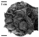
It has toe-beans!

Bring me the head of Arthropleura

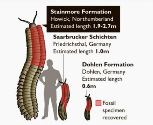
We’ve known about these amazing fossils from the lower Carboniferous for a while — it’s Arthropleura, a gigantic 2.5 meter long millipede. Imagine cleaning up your kitchen when a beast 2 or 3 times your length fluidly, sinuously crawls out from your baseboards. Wouldn’t that be neat?

One of the only problems with imagining that is that none of the fossils to date have had a head. Sure, it’s imposingly large, but what kind of face does it have? It’s a millipede, and millipedes are harmless detritivores who aren’t going to be a threat at all, unless you’re a pile of moldering leaves or a fungus. It’s centipedes that are primarily carnivores, with pointy sharp venomous forcipules that can deliver a nasty bite. That Arthropleura is in the millipede clade tends to blunt their potential menace.

Good news, time-traveling super-villains looking for a pet! The head of Arthropleura has at last been discovered, and it’s centipede-like, with strong bitey jaws, and also has stalked eyes. It’s a bit squished.

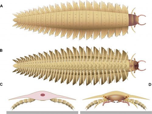
(A and B) Three-dimensional reconstruction. (A) Dorsal view. (B) Ventral view. (C and D) specimen inside the nodule. (C) Part. (D) Counterpart. Co, collum; DT, digestive tube; H, head; Pt, paratergite; S#, sternite number; St, syntergite; T#, tergite number; Te, telson. Reconstructions are made from Phoenix X-ray Phoenix V|tome|x CT scan. Scale bars, 1 cm (C and D) and 5 mm (A and B).

(A) Dorsal view. (B) Ventral view. (C) Back view. (D) Frontal view. Left maxillae were removed on (B) to better illustrate the mandible below. The red circle on (C) indicates the position of the digestive tract.

However, it’s still thought likely that it was a detritivorous. This has advantages for those of us who really want one as a pet: it’s still an intimidating creature, but in its free time it can roam the lair, cleaning up any untidiness.

Yes, I might fantasize a bit about keeping a few Arthropleura about the house. Better than a dog, anyhow.

These monsters are all dead

I hope you all like long tubular creatures, because that’s all I’ve got for you today. Maybe they’d be less horrifying if they had lots of legs?

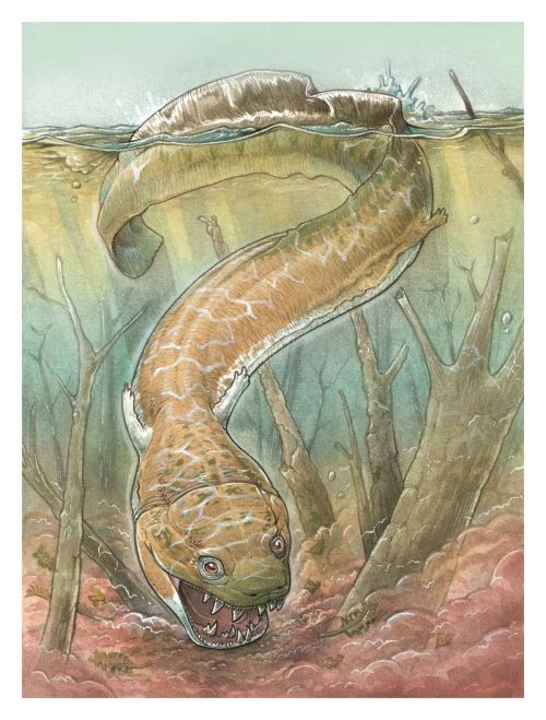
Here’s a 4-meter long salamander-like beast from the Permian, named Gaiasia.

I’ve seen giant salamanders before, but not ones with big box-like skulls full of razor-sharp fangs.

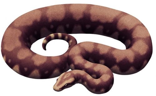
Here’s another muscular tube, Vasuki indicus, only 47 million years old, but somewhere around 10-15 meters long.

The amusing thing about this beast is that everyone in the popular press treatment is making it all about how long it is — it’s a partial skeleton, there’s not enough to determine exactly how long it is. It’s either shorter than Titanoboa, the gold standard of giant ancient snakes, or bigger than Titanoboa. It’s not a competition, people! They’re separated by about 10 million years. But of course they’re in competition for starring roles in cheesy sci-fi CGI epics.

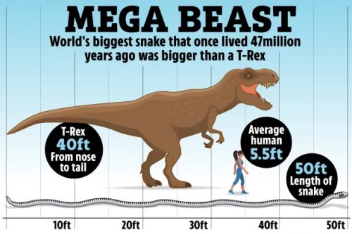
That’s why we’re seeing ridiculous comparisons like this one:

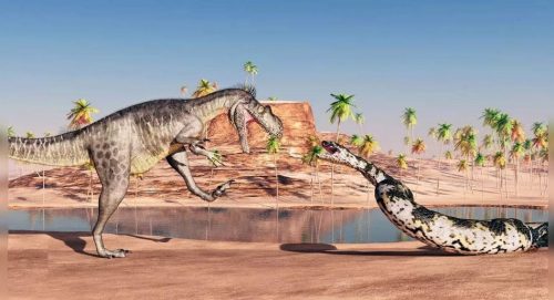
OK, the snake was longer than T. rex, but so what? It wasn’t as massive, and they were temporally distant from one another. This illustration reveals how some people are thinking:

That could be an ad for the next movie by The Asylum. These kinds of team-ups are popular to promote cheese, like Godzilla vs. Mechagodzilla or Dracula vs. Frankenstein. Learn to love Vasuki for itself, OK?

A Carboniferous arachnid

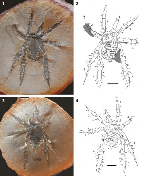
This week has been a good one for chelicerate evolution. Here’s another fossil, Douglassarachne acanthopoda, which was creeping around in the forests of Illinois in the late Carboniferous.

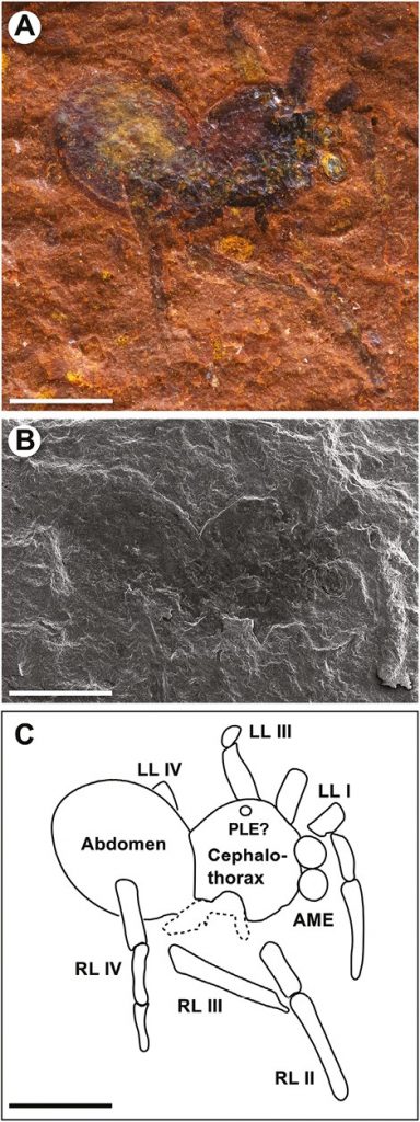
Douglassarachne acanthopoda n. gen. n. sp., holotype and only known specimen FMNH PE 91366; for interpretative drawings and scale, see Figure 2. (1) Part, detail of distal femur and more-distal podomeres, showing nature of curved macrospines on lateral edge of distal podomeres, bases of macrospines on dorsal surface of femur; (2) counterpart, detail of posterior opisthosoma showing bilobed structure at base of anal tubercle.

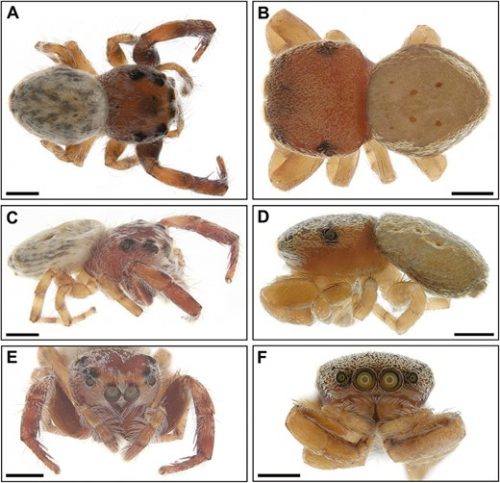
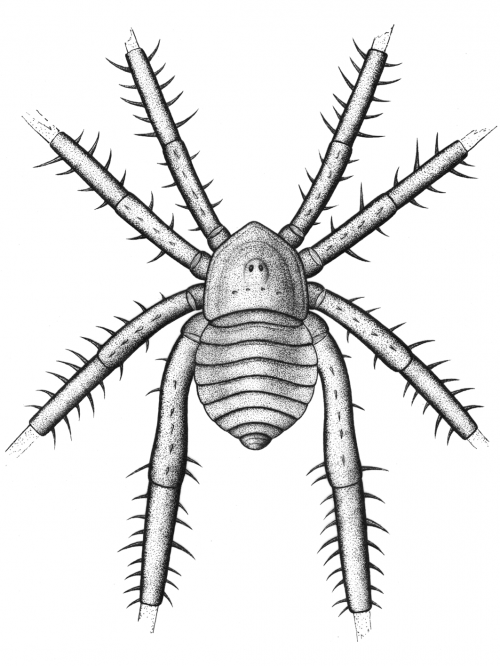
What is it? I don’t know. The authors are unsure. It’s an arachnid, but it could be in the spider lineage or the harvestman lineage, or it could be its own weird thing. It’s spiderish, anyway.

Douglassarachne acanthopoda n. gen. n. sp., reconstruction of the possible appearance of the animal in life.

An Ordovician ancestor to spiders

On this Memorial Day, I’m going to have to have a discussion with my spiders about their distinguished, noble ancestry. It was kind of Nature to publish a study of their many-times-great grand uncles, an ancient euchelicerate named Setapedites abundantis, a common fossil found in Moroccan sediments that are about 478 million years old, which puts it right in the middle of the Great Ordovician Biodiversification Event, a key moment in the evolution of modern taxa.

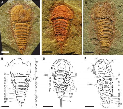
This is not a spider, though. It belongs in the euchelicerata, the large systematic group that includes spiders, scorpions, ticks, horseshoe crabs, sea scorpions, and other extinct groups. As you might guess from the name, a key feature is the presence of chelicerae, anterior appendages that in spiders carry the venomous fangs. It also has a common feature we see in both spiders and horseshoe crabs, the fusion of the anterior segments to form a prosoma, with posterior segments forming the abdomen or opisthosoma.

While it’s a cool looking little dude, it’s marine and pretty remote from modern chelicerates. From the dorsal side, it looks like an undistinguished little crustacean, of a type that was probably swarming in Ordovician seas.

A, B MGL.102899 and interpretative drawing, articulated specimen in dorsal view. C, D MGL.102828 and interpretative drawing, articulated specimen in dorsal view. E, F MGL. 102872 and interpretative drawing, articulated specimen in dorsal view. Abbreviations: btg, bipartite tergites; mr, median ridge; pl, pleura; pr, prosomal rim; saxn, sub-axial node; sr, sunken region; t1–11, tergites 1–11; t, telson; tk, telson keel. Scale bars, (A–F) 1 mm.

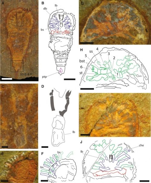
Where it gets interesting is when it’s flipped over, and you get a glimpse of the mass of limbs.

A, B YPM IP 517932c and interpretative drawing (counterpart), articulated specimen in ventral view. C, D YPM IP 517932c and interpretative drawing, chelicerae, and labrum anatomy detail. E, F Close-up of the prosoma of MGL.102934 and interpretative drawing, in dorso-lateral view. G, H Close-up of the prosoma of MGL.102634 and interpretative drawing, in ventral view. I, J Close-up of the prosoma of MGL.102800a under alcohol and polarized lighting, and interpretative drawing, in ventral view. Abbreviations: 1–6, podomeres 1–6 of the exopod; ptp, pretelsonic process; bs, basipodite; bst, brush-like setae; che, chelate podomere; db, doublure; lb, labrum; ss, single setae; st, pair of setae. Chelicerae are highlighted in gray, endopods in blue, exopods in green, opisthosomal appendages in red, and the pretelsonic process in purple. Scale bars, (A, B) 1 mm; (C, D) 100 µm; (E–K) 500 µm.

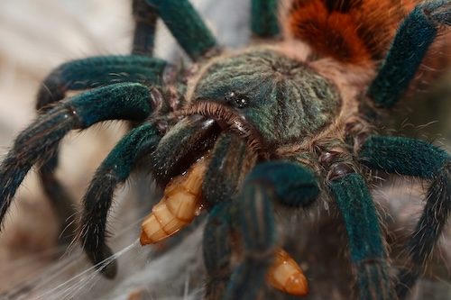
In front of the jaws proper (labrum, lb) it has a pair of small chelicerae (che). These have since evolved into the massive, sharptoothed chompers you can see my tarantula using to turn a mealworm into macerated mush.

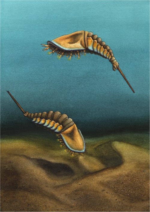
Setapedites wasn’t such a fierce predator. Here’s what it looked like.

Illustration by Elissa Sorojsrisom.

Cute, right? I don’t know why it’s drawn as a swimmer, though — with that anatomy, it looks more like a benthic organism.

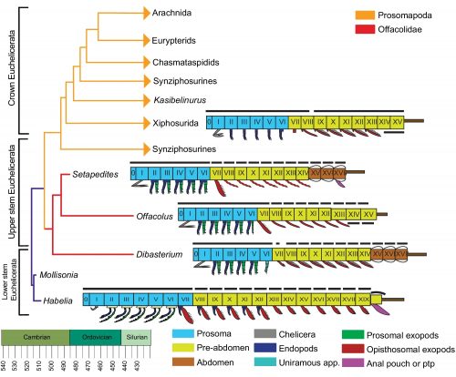
The final bit of interesting information is that they mapped out the correspondences in the segmentation of this animal with other, similar fossils and the extant Xiphosurians.

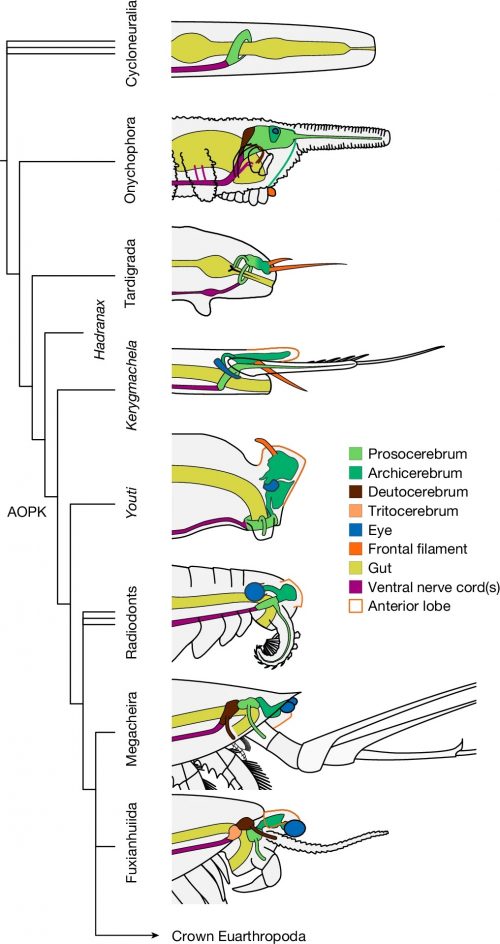
Simplified extended majority rule tree of a Bayesian analysis chronogram of euchelicerate relationships, based on a matrix of 39 taxa and 114 discrete characters, showing the position of Setapedites abundantis within Offacolidae. Lineages extending after the Silurian are indicated with arrowheads. Schematic models of the body organization in Habelia, Setapedites abundantis, Dibasterium, Offacolus, and Xiphosurida illustrate the origin and early evolution of euchelicerate uniramous prosomal appendages and tagmosis. Roman numbers designate somites. Prosoma somites are highlighted in blue, pre-abdomen somites in yellow, abdomen somites in brown, and the possible anal pouch or post-ventral structure (pvs) in purple. Black dorsal lines indicate tergites and cephalotorax. Schematic model of Xiphosurida Offacolus, and Dibasterium from 45, Habelia

Also of note: Setapedites had biramous appendages, a feature that is mostly kind of lost in modern arthropods — the outer branch got adapted into gills and lungs and even wings.

I can’t help but notice that domestication and artificial selection turns wolves in little yapping Pomeranians, but natural selection turns shrimp into tarantulas.

Burying the dead

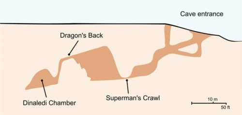
If you have a subscription to Netflix, you might want to watch Unknown: Cave of Bones, about the discovery of Homo naledi in the Rising Star cave system. It’s spectacular.

On the other hand, if you’re claustrophobic, you might want to skip it. I’m not particularly, but I watched the video of those women wriggling their way down a narrow crack to reach the Dinaledi Chamber gave me a rising sense of panic. There’s no way I could put myself in that position without having a screaming heebie-jeebie fit.

If you can get past that, though, it’s worth it to watch the adventure of science.

First feathers, now lips?

OK, guys, this has gone far enough. I grew up on images of dinosaurs that portrayed T. rex as hulking, scaly, snaggle-toothed dinosaurs, stomping through jungles and roaring. Now look at this…this…revisionism.

Theropod dinosaurs such as the iconic Tyrannosaurus rex have long been portrayed with their teeth fully visible, similar to extant crocodilians. This pattern of portrayal largely had to do with relatedness between dinosaurs and crocodilians and the relationship between tooth and jaw size. Cullen et al. tested hypothesized facial reconstruction in this group using histological analysis of tooth wear patterns and quantitative relationships between skull length and tooth size in both extinct and extant reptiles. Contrary to depictions that have dominated for more than a century, they found that theropods, including T. rex, had lips that covered their teeth, leaving them looking more like modern Komodo dragons than crocodiles.

Apparently, they covered up their dagger-like teeth behind lips, like perfect gentlemen.

Comparisons of the reconstructions of T. rex. (A) Skull, based on Field Museum of Natural History specimen FMNH PR 2081. (B to E) Two hypothetical flesh reconstructions, one with exposed teeth (B) and an associated cross section of the snout (C) and one with extraoral tissues covering the teeth (D) and an associated cross section of the snout (E).

I tell you, if some smarty-pants does an analysis next that shows that T. rex had a lovely singing voice and went “tweet tweet,” I’m going to turn this thing around and cancel the time machine project. There’ll be no point.

What an odd little beastie

I never heard of the Thylacocephala until I saw this video, and they are bizarre arthropods, now extinct, unfortunately. I learned something new!

At first I thought these were some strange planktonic creatures, but they were 20-30cm long. They were actively swimming predators that looked like some kind of remote drone submersible. They thrived from the Ordovician to the upper Cretaceous, making it kind of ridiculous that I knew nothing about them until now.

Vulcanized fossil spiders

And they fluoresce, too!

Part and counterpart of two fossil spiders shown in plain light and under UV illumination.

These are part of a well-known invertebrate fossil bed, 22.5 million years old, in France. It contains lots of well preserved insects and spiders, and one question is…why? What makes this particular place so good at preserving these delicate specimens?

The fluorescence was a clue. They dug into the chemistry of the fossils, and figured out that the glow was produced by the sulfurization of chitin, that as the dead spiders sank in the diatom-rich waters of an ancient pond, the sulfur in the diatoms reacted with the chitin carbohydrate to produce a tough carbon polymer, inedible to the microbes, that could last for millions of years.

Cartoon shows the entire proposed pathway: spider becomes entrained in planktonic diatom mat. Pieces of the diatom mat, both with and without spiders entrained within fall to the sediment floor against a background sedimentation of other diatoms and algae (gray dots). With time, these sediments become compressed and preserved into the rock record. a Chemical composition of chitin. Two chains of chitin are illustrated, organized in anti-parallel. Gray boxes indicate the carbonyl functionalities on the chitin. b Sulfonate-containing molecule, which are common in diatom EPS, can undergo microbial sulfate reduction (MSR), leading to the production of sulfide. c Chitin molecule after sulfurization. C–S bonds could potentially replace the carbonyl functionalities, and S–S bridges could form across the chitin chains. d Idealized molecule representing a chitin polymer after further diagenetic alteration, which could result in the formation of aromatized carbon.

I thought that was kind of neat. It’s also a reminder to biology students that you never know where that organic chemistry we make you take might be useful.