How to make a seahorse

Seahorses are weird animals. They depart from the typical streamlined torpedo shape of your average fish to construct this unusual twisted shape with dermal armor, toothless jaws, and a dependence on fins for propulsion — they’re just weirdos all around. How did they get to be this way?

One suggestion is that it is an extreme example of paedomorphosis, as presented in this paper: An embryonic arrest shapes the Syngnathid body plan: Insights from Seahorses, Pipefishes, and their Relatives.

The Syngnathidae (seahorses, seadragons, pipefish, pipehorses) exhibit a remarkable, enigmatic body plan, challenging conventional explanations for their fused jaws, toothlessness, cartilaginous skeleton, fin loss, male pregnancy, and their distinctive morphology, which includes the acute head-trunk angle of seahorses and the family’s unique curling, often prehensile, tail. We propose a unifying, parsimonious hypothesis, termed “pharyngulation,” that the entire lineage originated from a profound paedomorphic arrest (retention of juvenile traits) during a specific embryonic pharyngula stage. This arrest, likely driven by ancestral Hox gene cluster disintegration, fundamentally halted morphological progression in a common teleost ancestor. This single event explains their entire suite of primary characteristics–including universal low body mass and volume and unique A-P locomotion. It also establishes a framework to differentiate these foundational family-defining traits from ancestral features shared with the broader Syngnathiformes order (such as the elongated snout, as exemplified by Trumpetfish) and from later adaptive refinements, such as the leaf-like appendages in seadragons. Our “pharyngulation” hypothesis offers a novel, testable model for macroevolutionary innovation, demonstrating how a singular, profound alteration to a conserved developmental program can rapidly forge a new, viable body plan. This concept, synthesizing evidence from genomics, the fossil record, and developmental biology, is of broad interest to evolutionary biologists and developmental biologists alike.

Unfortunately, this paper only presents a hypothesis — no methods, no experiments, no substantial comparative data. I’ll forgive that since it does introduce the term “pharyngulation” into the scientific literature.

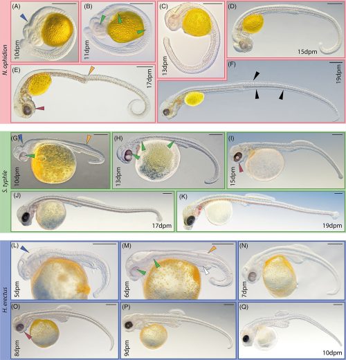
I was provoked to dig a little deeper, and found this paper: A comparative analysis of the ontogeny of syngnathids (pipefishes and seahorses) reveals how heterochrony contributed to their diversification. It supports some of the ideas of the first paper — heterochrony is right there in the title — and also includes some beautiful photos of syngnathid embryos.

Segmentation and early organogenesis development in examined syngnathids. Nerophis ophidion (A-F), Syngnathus typhle (G-K), and Hippocampus erectus (L-Q), respectively. In this period, species-specific characteristics develop more clearly. Arrowheads: blue = hind brain vesicle, green = pigmentation, rufous = mandibular arch, orange = dorsal fin condensations, white = hypertrophic hindgut, black = fin fold. Scale is 500 μm; dpm = days post mating

That’s a stage close to what we’d call the pharyngula stage (which doesn’t have a single discrete marker), and they look familiar — they look like longer, skinnier, more slowly developing zebrafish embryos, where the 19 day syngnathid looks like a 19 hour zebrafish. We have to wait a week or more to see an embryo that is comparable to, but very different from, a 24-48 hour old zebrafish embryo.

Organogenesis to release development in examined syngnathids. Nerophis ophidion (A-D), Syngnathus typhle (E-I), and Hippocampus erectus (J-O), respectively. The last prerelease period is characterized by snout elongation, continued pigmentation and the conclusion of allometric fin outgrowth. Arrowheads: black = fin fold. Scale is 500 μm; dpm = days post mating

And that’s where I see the problem with the paedomorphosis explanation. This is not simply a case of developmental arrest. There are clear differences in growth prior to the pharyngula stage, and the pharyngula stage is, at best, a point of divergence in development, and so much of what is happening at that point and thereafter is the appearance of evolutionary novelties. It’s not so much that the pattern stops, as that there are a whole host of additions to the organization of the syngnathid body plan in embryogenesis.

Also, data is always pretty.

A familiar story told again

The pre-Cambrian animal rollover: A micro-evolutionary event becomes a major macro-evolutionary distinction in 600 million years.

At 10am Central tomorrow morning, you can watch this video about the dorsal-ventral inversion of key signaling molecules in the developing embryo. Or you can watch it on Patreon right now if you’re a member.

This is a discussion of this paper:
De Robertis EM, Tejeda-Munoz N (2022) Evo-Devo of Urbilateria and its larval forms. Developmental Biology 487:10-20.

A spider with two hoo-has

Nice.

Almafuerte peripampasica, female. A, prosoma and opisthosoma (dorsal view). B, opisthosoma (ventral view, arrow pointing to the duplicated epigyne). Note the difference in size and the slightly asymmetry. C, normal epigyne (arrow pointing to larva). D, duplicated epigyne. Specimen locality: Argentina: Córdoba: Parque Nacional Quebrada del Condorito, “sitio 2”, -31.63481, -64.71087, alt. 1846m, M. Izquierdo, D. Abregú, C. Mattoni, col. Sep. 16, 2019, under stones. LABRE-Ar 498, voucher MAI-4754.

Developmental variations always catch my eye, and this one is interesting from the standpoint of looking for spatial specifications. The normal blueprint for the spider body plan puts the epigyne at the anterior end of the abdomen, but here’s one way back near the spinnerets. What triggered formation at that position? It’s also non-functional, they think — I’d want to know more about the internal organs, what’s connected to what.

In general, malformations in spiders involve chelicerae, pedipalps, walking legs, and eyes, but those affecting female reproductive structures are not so frequent. A teratological case of a spider with a duplicated reproductive structure is described. The female specimen has the typical epigyne and a second one near the spinnerets. The second epigyne is less developed and seems to be non-functional. Similar malformations have been reported for Amaurobiidae and Salticidae, and here is presented for Gnaphosidae. Although it is widely known that temperature and humidity may induce abnormalities under experimental conditions, the causes behind teratological genitalia in wild females are unknown. This case opens the question of the origin of such a malformation and the ontogeny of female reproductive organs in spiders.

Also, that’s an adult of respectable size, so the ectopic organ doesn’t seem to have affected its viability. I’m going to have to spend some time looking up spiders’ skirts this summer.

Sex combs!

I mentioned sex combs a while back, so I thought I’d clarify a bit — I hope none of you rushed out to buy one for yourself (I don’t think human sex combs exist, but if they do, I don’t need to know.) Sex combs are secondary sexual characteristics found in on the forelegs of only male Drosophilidae. They are small dark patches of bristles on the tarsus of the first leg, and they are not something you’d notice if you saw a fly buzzing in your kitchen — you have to knock them out and carefully scrutinize the limbs with a hand lens or microscope to see them, but they’re important for recognizing the sex of a fly definitively. I tell my genetics students that you can tell the sexes apart by the shape of the abdomen or the pigment patterns, but to be really sure you should check for the presence or absence of the sex combs.

Sex comb in Drosophila melanogaster male: a) front leg with sex comb marked with black arrow; b) sex comb bristles

They don’t look like much, but they also matter to female flies. Mutants or surgically modified male flies with the sex combs reduced suffer with lower reproductive success. The flies use them as gentle grasping tools to separate her wings, grasp her abdomen, and tease open the genitals, so of course they’re subject to selection. Different species of fruit flies exhibit different patterns of sex combs, and we observe natural variation within a species.

Sex comb (SC) diversity. A phylogeny of eight comb-bearing species, assuming that the montium subgroup is a sister-taxon to the
Oriental lineage, as in Kopp (2006). Branch lengths are not to scale. Note the variation in the length, size and orientation of the SCs.

Variation within and between species makes sex combs of great interest to evolutionary biologists. Here’s an illustration of the variation we can see.

Variation in sex comb tooth number and development. (A–C) Exam-
ples of Drosophilidae forelegs with long combs. (D and E) D. melanogaster
forelegs. (F–J) Schematics of foreleg development of the top Drosophila legs.
(K–O) Examples of
D. melanogaster perturbations in sex comb tooth number.
(A–C and F–H) Drosophila species with long sex combs achieve vertical orien-
tation by different mechanisms: (i) Teeth initially form in a vertical orientation
(F) (e.g., D. ficusphila t1–t2); (ii) rotation of a long row (G) (e.g., D. guanche t1–t2
and D. rhopaloa t1); (iii) rotation of multiple small rows and posterior fusion
into a long sex comb (H) (e.g., D. rhopaloa t2). (D, E, I, and J) In D. melanogaster, the male sex comb rotates from a horizontal to a vertical position
(diagrammed in D), while TRs remain horizontal. The only exception is the
most distal transverse row (red dotted box in D and I), which bends proximally
close to the top part of the sex comb. In contrast, the female rows of bristles
homologous to the sex comb remain static during development (brackets in E
and J). In order to study the phenotypic and developmental effect of changing
the number of sex comb teeth, this trait was perturbed using artificial selection
(K and L), mutants (M–O), and UAS-Scr RNAi transgenic lines . Gray circles represent sex comb teeth in the initial position and
black circles represent sex comb teeth in the final position. Empty circles represent the TR bristles. Gray arrows indicate movement of individual tooth or
sex comb rows. Red brackets indicate sex combs or homologous female bristles. Red numbers represent the range of sex comb teeth in each line. babPR72, bric à bracPR72; scd, sex combs distal; Scr, sex combs reduced; t1 and t2, first and
second tarsal segment, respectively. Distal is down and posterior is to the right.
Scale bar: 20 μm.

Focus for now on the top panel on the right, which shows a male and a female foreleg. They both have hairy legs, and in the default pattern seen in the females is a series of bristles in transverse rows (TRs) in arrays marching down the leg. The flies specifically use these bristles to groom their eyes — if you look closely, flies are remarkably tidy and neat.

The TRs are illustrated diagrammatically as small open circles in rows, the base pattern. These bristles are also developmentally interesting, because the way you make a sex comb is you express a set of specific genes in the distal two rows, and the whole structure rotates 90° to form a longitudinal comb. This opens up a whole set of informative interactions — the rotation is essential for function, and is subject to constraints imposed by adjacent tissues. I’ve been reading papers for the past week focused on the developmental and evolutionary significance of this tiny, odd, little known structure in flies. You should read some of these papers, too! I’m a sucker for anything evo-devo, and that’s what this little patch of hairs illustrates.

The most complex and diverse secondary sexual character in Drosophila is the sex comb (SC), an arrangement of modified bristles on the forelegs of a subclade of male fruit flies. We examined SC formation in six representative nonmodel fruit fly species, in an effort to understand how the variation in comb patterning arises. We first compared SC development in two species with relatively small combs, Drosophila takahashii, where the SCs remain approximately transverse, and Drosophila biarmipes, where two rows of SC teeth rotate and move in an anterior direction relative to other bristle landmarks. We then analyzed comb ontogeny in species with prominent extended SCs parallel to the proximodistal axis, including Drosophila ficusphila and species of the montium subgroup. Our study allowed us to identify two general methods of generating longitudinal combs on the tarsus, and we showed that a montium subgroup species (Drosophila nikananu) with a comb convergently similar in size, orientation and position to the model organism Drosophila melanogaster, forms its SC through a different developmental mechanism. We also found that the protein product of the leg patterning gene, dachshund (dac), is strongly reduced in the SC in all species, but not in other bristles. Our results suggest that an apparent constraint on SC position in the adult may be attributable to at least two different lineage-specific developmental processes, although external forces could also play a role.

Atallah J, Liu NH, Dennis P, Hon A, and Larsen EW (2009) Developmental constraints and convergent evolution in Drosophila sex comb formation. Evolution & Development 11(2): 205-218.

Malagón JN, Ahujab A, Sivapatham G, Hung J, Leea J, Muñoz-Gómez SA, Atallah J, Singh RS, and Larsena E (2013) Evolution of Drosophila sex comb length illustrates the inextricable interplay between selection and variation www.pnas.org/cgi/doi/10.1073/pnas.1322342111

Another step in the evolution of multicellularity

I’m not a fan of phys.org — they summarize interesting articles, but it’s too often clear that their writers don’t have a particularly deep understanding of biology. I wonder sometimes if they’re just as bad with physics articles, and I just don’t notice because I’m not a physicist.

Anyway, here’s a summary that raised my hackles.

Chromosphaera perkinsii is a single-celled species discovered in 2017 in marine sediments around Hawaii. The first signs of its presence on Earth have been dated at over a billion years, well before the appearance of the first animals.

A team from the University of Geneva (UNIGE) has observed that this species forms multicellular structures that bear striking similarities to animal embryos. These observations suggest that the genetic programs responsible for embryonic development were already present before the emergence of animal life, or that C. perkinsii evolved independently to develop similar processes. In other words, nature would therefore have possessed the genetic tools to “create eggs” long before it “invented chickens.”

First two words annoyed me: Chromosphaera perkinsii ought to be italicized. Are they incapable of basic typographical formatting? But that’s a minor issue. More annoying is the naive claim that a specific species discovered in 2017 has been around for a billion years. Nope. They later mention that it might have “evolved independently to develop similar processes”, which seems more likely to me, given that they don’t provide any evidence that the pattern of cell division is primitive. It’s still an interesting study, though, you’re just far better off reading the original source than the dumbed down version on phys.org.

All animals develop from a single-celled zygote into a complex multicellular organism through a series of precisely orchestrated processes. Despite the remarkable conservation of early embryogenesis across animals, the evolutionary origins of how and when this process first emerged remain elusive. Here, by combining time-resolved imaging and transcriptomic profiling, we show that single cells of the ichthyosporean Chromosphaera perkinsii—a close relative that diverged from animals about 1 billion years ago—undergo symmetry breaking and develop through cleavage divisions to produce a prolonged multicellular colony with distinct co-existing cell types. Our findings about the autonomous and palintomic developmental program of C. perkinsii hint that such multicellular development either is much older than previously thought or evolved convergently in ichthyosporeans.

Much better. The key points are:

  • C. perkinsii is a member of a lineage that diverged from the line that led to animals about a billion years ago. It’s ancient, but it exhibits certain patterns of cell division that resemble those of modern animals.
  • Symmetry breaking is a simple but essential precursor to the formation of different cell types. The alternative is equipotential cell division, one that produces two identical cells with equivalent cellular destinies. Making the two daughter cells different from each other other opens the door to greater specialization.
  • Palintomic division is another element of that specialization. Many single-celled organisms split in two, and each individual begins independent growth. Palintomic division involves the parent cell undergoing a series of divisions without increasing the total cell volume. They divide to produce a pool of much smaller cells. This is the pattern we see in animal (and plant!) blastulas: big cell dividing multiple times to make a pile of small cells that can differentiate into different tissues.
  • Autonomy is also a big deal. They looked at transcriptional activity to see that daughter cells had different patterns of gene activity — some cells adopt an immobile, proliferative state, while others develop flagella and are mobile. This is a step beyond forming a simply colonial organism, is a step on the path to true multicellularity.

Cool. The idea is that this organism suggests that single-celled organisms could have acquired a toolkit to enable the evolution of multicellularity long before their descendants became multicellular.

I have a few reservations. C. perkinsii hasn’t been sitting still — it’s had a billion years to evolve these characteristics. We don’t know if they’re ancestral or not. We don’t get any detailed breakdown of molecular homologies in this paper, so we also don’t know if the mechanisms driving the patterns are shared.

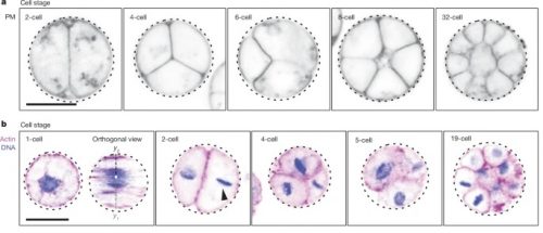
I was also struck by this illustration of the palintomic divisions the organism goes through.

a, Plasma membrane-stained (PM) live colonies at distinct cell stages, highlighting the patterned cleavage divisions, tetrahedral four-cell stage and formation of spatially organized multicellular colonies (Supplementary Video 5). b, Actin- (magenta) and DNA-stained (blue) colonies at distinct cell stages showcasing nuclear cortical positioning, asymmetrical cell division (in volume and in time) and the formation of a multicellular colony. This result has been reproduced at least three independent times.

Hang on there : that’s familiar. D’Arcy Wentworth Thompson wrote about the passive formation of cell-like cleavage patterns in simple substrates, like oil drops and soap bubbles, in his book On Growth and Form, over a century ago. You might notice that these non-biological things create patterns just like C. perkinsii.

Aggregations of oil-drops. (After Roux.) Figs. 4–6 represent successive changes in a single system.

Aggregations of four soap-bubbles, to shew various arrangements of the intermediate partition and polar furrows.

An “artificial tissue,” formed by coloured drops of sodium chloride solution diffusing in a less dense solution of the same salt.

That does not undermine the paper’s point, though. Multicellularity evolved from natural processes that long preceded the appearance of animals. No miracles required!

Wilson’s Principles of Teratology

It’s another busy week of EcoDevo, and even though the campus was closed I still had to give a lecture on endocrine disruptors. I started by laying out Wilson’s Principles of Teratology…wait, what? You don’t know them? I guess I’d better explain them to the internet at large.

These principles are a bit like Koch’s Principles, only for teratology — you better know them if you want to figure out the causes of various problems at birth, and you do: about 3% of all human births express a defect serious enough for concern. Here’s the list:

  1. Susceptibility to teratogenesis depends on the genotype of the conceptus and the manner in which this interacts with adverse environmental factors.
  2. Susceptibility to teratogenesis varies with the developmental stage at the time of exposure to an adverse influence. There are critical periods of susceptibility to agents and organ systems affected by these agents.
  3. Teratogenic agents act in specific ways on developing cells and tissues to initiate sequences of abnormal developmental events.
  4. The access of adverse influences to developing tissues depends on the nature of the influence. Several factors affect the ability of a teratogen to contact a developing conceptus, such as the nature of the agent itself, route and degree of maternal exposure, rate of placental transfer and systemic absorption, and composition of the maternal and embryonic/fetal genotypes.
  5. There are four manifestations of deviant development (death, malformation, growth retardation and functional defect).
  6. Manifestations of deviant development increase in frequency and degree as dosage increases from the No Observable Adverse Effect Level (NOAEL) to a dose producing 100% lethality (LD100).

The first two tell you what is tricky about teratology. There are multiple variables that affect the response: genetic variability in the conceptus (and, I would suggest, maternal variations), and also timing is critical. A drug might do terrible things to an embryo at 4 weeks, but at 3 months the fetus shrugs it off.

Ultimately, though, the teratogen is having some specific effect (3) on a developing tissue. We just have to figure out what it is, while keeping in mind that that effect might be hiding in a maze of genetics (1) and time (2).

Another complication is that in us mammals the embryo is sheltered deep inside the mother, who has defense mechanisms. The agent has to somehow get in (4). A complication within a complication: sometimes the teratogenic agent is harmless until Mom chemically modifies it as part of her defense, and instead creates a more potent poison.

#5 is just listing the terrible outcomes of screwing with development.

#6 I do not trust. It’s saying the effect is going to follow a common sense increase with increasing dosage, but even that isn’t always true. There is a phenomenon called the inverted-U response where the effect increases with dosage, then plateaus, and then drops off at high concentrations. We’re dealing with complex regulatory phenomena with multiple molecular actors that may have unpredictable interactions. There are teratogens that do terrible things to embryos at low concentrations, but do nothing at ridiculously high concentrations — as if the high dose triggers effective defense mechanisms that the low dose sidesteps.

I had to review these principles in class yesterday, because although I’d also discussed them earlier in the semester, we are currently dealing with teratogens of monstrous subtlety, these compounds that mimic our own normal developmental signals, the same signals our bodies use to assemble critical organ systems. It’s as if some joker were placing inappropriate traffic signals along a busy highway — most would do no harm, but some may totally confuse travelers who then end up detouring up into the kidneys rather than down the genitals, as they preferred, or they end up crashing into the thyroid.

Unfortunately, in this case the responsible jokers are mainly gigantic megacorporations who are spewing these dangerous signals all over the countryside…and then we get to wait until the people swimming in them try to have children, and then the teratologists get to say “death, malformation, growth retardation and functional defect”.


In case you were wondering, Wilson didn’t come up with his list first — a 19th century scientist named Gabriel Madeleine Camille Dareste did it first. No, not first. Lots of people have been documenting these developmental problems as long as there’s been writing, like on this Chaldean tablet:

When a woman gives birth to an infant:
With the ears of a lion There will be a powerful king
That wants the right ear The days of the king will be prolonged
That wants both ears There will be mourning in the country
Whose ears are both deformed The country will perish and the enemy rejoice
That has no mouth The mistress of the house will die
Whose nostrils are absent The country will be in affliction and the house of the man will be ruined
That has no tongue The house of the man will be ruined
That has no right hand The country will be convulsed by an earthquake
That has no fingers The town will have no births
That has the heart open with no skin The country will suffer from calamities
That has no penis The master of the house shall be enriched by the harvest of his field
Whose anus is closed The country shall suffer from want of nourishment
Whose right foot is absent His house will be ruined and there will be abundance in that of the neighbor
That has no feet The canals of the country will be cut and the house ruined
If a queen gives birth to:
An infant with teeth already cut The days of the king will be prolonged
A son and a daughter at the same time The land will be enlarged
An infant with the face of a lion The king will not have a rival
An infant with 6 toes on both feet The king shall rule the enemies’ country

Nowadays we’re more interested in causes than imagined consequences, I hope.

You know, alcohol is not good for children and other growing things

A few weeks ago, I had an absolutely delicious stout at a brew pub in Alexandria. I’m going to have to remember it, because it may have been the last time I let alcohol pass these lips. Why? Because I’m slowly turning into one of those snooty teetotalers who tut-tut over every tiny sin. It started with vegetarianism, now it’s giving up alcohol, where will it end? Refusing caffeine, turning down the enticements of naked women, refusing to dance? The bluenose in me is emerging as I get older. I shall become a withered, juiceless old Puritan with no joy left in me.

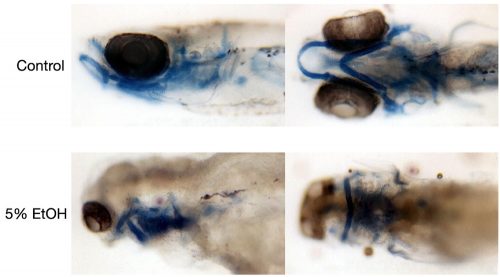
It didn’t help that last week I was lecturing on alcohol teratogenesis in my eco devo course, and it was reminding me of what a pernicious, sneaky molecule it is. I’ve known a lot of this stuff for years, but there’s a kind of blindness brought on by familiarity that led me to dismiss many of the problems. You know the phenomenon: “it won’t affect me, I only drink in moderation” and other excuses. Yeah, no. There are known mechanisms for how alcohol affects you, besides the obvious ones of inebriation.

  1. It induces cell death.
  2. It affects neural crest cell migration.
  3. It downregulates sonic hedgehog, essential for midline differentiation.
  4. It downregulates Sox5 and Ngn1, genes responsible for neuron growth and maturation.
  5. It weakens L1-modulated cell adhesion.

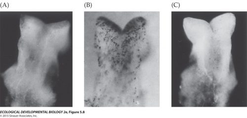
I already knew all about those first four — I’ve done experiments in zebrafish like these done in mice.

Take a normal, healthy embryo like the one in A, expose it to alcohol, and stain the brain for cell death with any of a number of indicator dyes, like Nile Blue sulfate in this example B (I’ve used acridine orange, it works the same way). That brain is speckled with dead cells, killed by alcohol. If you do it just right, you can also see selective cell death in neural crest cell populations, so you’re specifically killing cells involved in the formation of the face and the neurons that innervate it. In C, you can see the rescuing effects of superoxide dismutase, a free radical scavenger, and that tells you that one of the mechanisms behind the cell death is the cell-killing consequences of free radicals. I could get a similar reduction in the effects with megadoses of vitamin C, but that doesn’t mean a big glass of orange juice will save you from your whisky bender.

I was routinely generating one-eyed jawless fish, a consequence of the double-whammy of knocking out sonic hedgehog and cell death in the cells that make branchial arches.

You can wave away these results by pointing out those huge concentrations of alcohol we use to get those observable effects, but we only do that because we don’t have the proper sensitivity to detect subtle variations in the faces of mice or fish. So we crank up the dosage to get a big, undeniable effect.

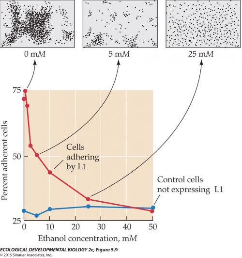
I only just learned about the L1 effects, and that’s a case where we have a sensitive assay for alcohol’s effects. L1 is a cell surface adhesion molecule — it helps appropriate populations of cells stick together in the nervous system. It also facilitates neurite growth. It’s good for happy growing brains.

It also makes for a relatively easy and quantitative assay. Put neuronal progenitors that express L1 in a dish, and they clump together, as they should in normal development. Add a little alcohol to the medium, and they become less sticky, and the clumps disperse.

What’s troubling about this is the dosage. Adhesion is significantly reduced at concentration of 7mM, which is what the human blood alcohol level reaches after a single drink. The fetal brain may not be forming as robustly when Mom does a little social drinking that doesn’t leave her impaired at all, not even a slight buzz.

Maybe you console yourself by telling yourself a little bit does no harm, your liver soaks up most of the damage (and livers are self-repairing!), that it’s only binge drinkers who have to worry about fetal alcohol syndrome, etc., etc., etc. We have lots of excuses handy. Humans are actually surprisingly sensitive to environmental insults, we have mechanisms to compensate, but there’s no denying that we’re modifying our biochemistry and physiology in subtle ways by exposure to simple molecules.

Now maybe you also tell yourself that you’re a grown-up, I’m talking about fetal tissues, and you also don’t intend to get pregnant in the near future or ever. I’m also a great big fully adult person who is definitely not ever going to get pregnant, but development is a life-long process, and we’re all fragile creatures who nonetheless soak up all kinds of interesting and dangerous chemicals during our existence. We know alcohol will kill adult brain cells, but what else does it do? Do you want to be a guinea pig? I think that, as I age, I am becoming increasingly aware of all the bad stuff I did to myself in my heedless youth, and am starting to think that maybe I need to be a little more careful, belatedly.

Oh, you want some reassuring information? Next week we’re discussing endocrine disruptors in my class — DDT, DES, BPA, PCB, etc. — all these wonderful products of plastics and petrochemical technology. You’re soaking in them right now. They never go away. How’s your sperm count looking? Any weird glandular dysplasias? Ethanol looks pretty good compared to chlorinated and brominated biphenyls.

Grow a segmented spine!

You want to see something awesome? This is a video of human embryonic cells developing in a dish and activating the segmentation clock to build a series of somites.

That embryos do this is pretty neat but not surprising. It’s easy to see that process going on in zebrafish embryos, where a new segmental blob pinches off every 20 minutes. What’s cool is that they’ve pared down the system to just what looks like presomitic mesoderm — no gut endoderm, no nervous system or ectoderm, just those mesodermal cells grouping and organizing to make pre-muscle tissue. At first, I thought, no big deal. We’ve observed vertebrate segmentation in fish and frogs and chicks for many decades, why would we need to look at it in human embryos? They almost certainly use the same molecules and interactions as mice or any other vertebrate. It’s got to be using Notch signaling, after all.

What’s special here, though, is that they’ve reduced the complexity of the system to a remarkable degree. You don’t need a nervous system or a gut or even, it seems, a notochord to assemble segmental organization. They’re even getting colinear expression of HOX genes! That’s the surprise to me, that so little is required to trigger oscillatory expression and patterning. And that’s the point the authors emphasize.

Our bottom-up experimental approach demonstrates that complex developmental events such as somitogenesis, can be deconstructed and dissected into discrete “building blocks” of developmental principles which are usually intricately connected and cannot be easily uncoupled in vivo. Axioloids, a self-organizing in vitro model of human axial development allowed us to individually assess and manipulate such building blocks at the molecular, cellular and morphogenetic level. Axioloids -a surrogate model of the human embryonic tail and forming axis- are capable of recapitulating core features of human somitogenesis, and represent an exciting new platform to investigate axial development and disease in a human context.

Don’t read the YouTube comments. There are people complaining that it’s a human life with a unique genetic code (no, it’s not a person, and these embryonic cultures cannot develop into a person — while scraps of mesoderm might be enough to make somites, it’s not sufficient to build a whole human being) and Stop trying to PLAY GOD!!! (this isn’t god-like, it’s biology).

Bad science tries to drip its way into everything

You want to read a really good take-down of a bad science paper? Here you go. It’s a plea to Elsevier to retract a paper published in Personality and Individual Differences because…well, it’s racist garbage, frequently cited by racists who don’t understand the science but love the garbage interpretation. It really is a sign that we need better reviewers to catch this crap.

The paper is by Rushton, who polluted the scientific literature for decades, and Templer, published in 2012. It’s titled “Do pigmentation and the melanocortin system modulate aggression and sexuality in humans as they do in other animals?”, and you can tell what it’s trying to do: it’s trying to claim there is a genetic linkage between skin color and sexual behavior and violence, justifying it with an appeal to biology. It fails, because the authors don’t understand biology or genetics.

They’re advocating something called the pleiotropy hypothesis, which is the idea that every gene has multiple effects (this is true!), and that therefore every phenotype has effects that ripple across to every other phenotype (partially, probably mostly true), so that seeing one aspect of a phenotype means you can make valid predictions about other aspects of the phenotype (mostly not at all true). This allows them to abuse a study in other mammals to claim that human outcomes are identical. Here’s the key graf:

The basis of the pleiotropy hypothesis presented by Rushton and Templer hinges on a citation from Ducrest et al. (2008), which posits ‘pleiotropic effects of the melanocortins might account for the widespread covariance between melanin-based coloration and other phenotypic traits in vertebrates.’ However, Rushton and Templer misrepresent this work by extending it to humans, even though Ducrest et al. (2008) explicitly state, ‘these predictions hold only when variation in melanin-based coloration is mediated by variation in the level of the agonists at MC1R… [conversely] there should be no consistent association between melanin-based coloration and other phenotypic traits when variation in coloration is due to mutations at effectors of melanogenesis such as MC1R [as is the case in humans].’ Ducrest et al. continue, ‘variation in melanin-based coloration between human populations is primarily due to mutations at, for example, MC1R, TYR, MATP and SLC24A5 [29,30] and that human populations are therefore not expected to consistently exhibit the associations between melanin-based coloration and the physiological and behavioural traits reported in our study’ [emphasis mine]. Rushton and Templer ignore this critical passage, saying only ‘Ducrest et al. (2008) [caution that], because of genetic mutations, melanin-based coloration may not exhibit these traits consistently across human populations.’ This is misleading. The issue is not that genetic mutations will make melanin-based pleiotropy inconsistent across human populations, but that the genes responsible for skin pigmentation in humans are completely different to the genes Ducrest et al. describe.

To translate…developmental biologists and geneticists are familiar with the concept of an epistatic pathway, that is, of genes affecting the expression of other genes. So, for instance, Gene A might switch on Gene B which switches on Gene C, in an oversimplified pattern of regulation.

Nothing is ever that simple, we know. Gene A might also switch on Gene Delta and Gene Gamma — this is called pleiotropy, where one gene has multiple effects. And Gene Gamma might also activate Gene B, and Gene B might feed back on Gene A, and B might have pleiotropic effects on Gene Beta and Gene E and Gene C.

This stuff gets delightfully tangled, and is one of the reasons I love developmental biology. Everything is one big complex network of interactions.

What does this have to do with Rushton & Templer’s faulty interpretation? They looked at a study that identified mutations in a highly pleiotropic component of the pigmentation pathway — basically, they’re discussing Gene A in my cartoon — and equating that to a terminal gene in humans, equivalent to Gene C in my diagram. Human variations in skin color are mostly due to mutations in effector genes at the end of the pathway, like MC1R. It will have limited pleiotropic effects compared to genes higher up in the epistatic hierarchy, like the ones Ducrest et al. described. Worst of all, Ducrest et al. explicitly discussed how the kind of comparison Rushton & Templer would make is invalid! They had to willfully edit the conclusions to make their argument, which is more than a little dishonest.

It reminds me of another recent disclosure of a creationist paper that also misrepresented its results. This paper, published in the International Journal of Neuroscience, openly declared that it had evidence for creationism.

In the paper, Kuznetsov reportedly identified an mRNA from one vole species that blocked protein synthesis in a related vole species. That same mRNA, however, did not block translation in the original vole species or another species that was more distantly related. The finding, Kuznetsov wrote in his report, supported “the general creationist concept on the problems of the origin of boundless multitudes of different and harmonically functioning forms of life.”

I vaguely remember reading that paper and rolling my eyes at how weak and sloppy the data was — it was never taken seriously by anyone but creationists. I don’t recall the details, though, because it was published 30 years ago, and is only now being retracted, after decades of the author fabricating data and being so obvious about it that he was fired as editor of two journals in 2013. The guy had a reputation, shall we say. Yet he managed to maintain this academic facade for years.

Phillipe Rushton had similarly managed to keep up the pretense of being a serious academic for an awfully long time, right up until his death in 2012. He used his reputation to spray all kinds of fecal nonsense into the scientific literature, and that’s why you have to maintain a skeptical perspective even when reading prestigious journals.