I went through high school thinking that evolution was pretty neat. I read all the popular treatments — those Time/Life books, National Geographic, science magazines, etc. — and they made it sound like Darwin had got everything right in 1859. I could go along with that.
But then, in 1975, I went off to college and had access to a university library and started reading the real hardcore stuff. I was just mainlining PNAS & Science & Nature & Evolution & the Journal of Embryology and Experimental Morphology and soaking in all this exciting stuff, and that’s when I discovered the concept of genetic drift (specifically, thanks to Lewontin). My mind was blown. Suddenly, questions I had never even thought to ask were answered with clarity. Certainly nothing in biology makes sense except in the light of evolution, but likewise nothing in evolution makes sense except in the light of neutral genetic drift.
I guess Holly Dunsworth had the same revelation, and also noticed the sad gap in public education.
In the USA, K-12 evolution education standards are missing GENETIC DRIFT as well as the word NEUTRAL.
(see here: https://www.nextgenscience.org/topic-arrangement/hsnatural-selection-and-evolution)
I don’t know how to understand life’s perpetual change without those fundamentals.
If students know about meiosis (which *is* in the standards), then they know about genetic drift. It’s just a matter of linking meiosis to evolution.
Genetic drift is a very simple concept and makes natural selection make a whole lot more sense!
I shout about this on Twitter and I get “just be happy anyone teaches evolution in K-12 at all” and “teachers don’t know about drift” in response. But evolution without genetic drift is not evolution, and if teachers knew about drift, then they’d be more comfortable teaching evolution. I guarantee it!
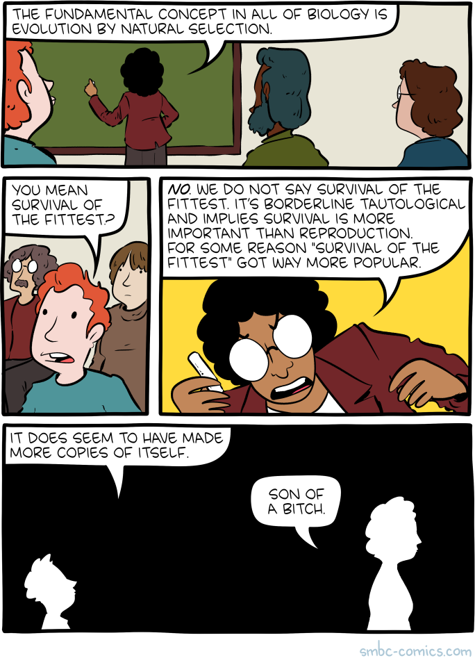
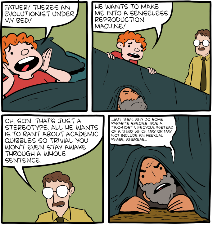
Without drift, it’s too easy to just replace God with natural selection. And that habit of narrating evolution by giving agency to natural selection is super-duper weird for non-believers let alone for believers! (This a no hatred of religion, faith, or creationism zone.)
Genetic drift paves the way for thinking about and then narrating evolution as the constant change that life/nature/biology is. Everyone gets that time is just constant change and they will get that nature/biology/life is too. They embody it themselves, being different from their parents.
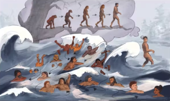
Plus, continuing to teach evolution as only being natural selection (which is what the standards are doing), is also dangerous. That selection-obsessed mindset is tied to racism, sexism, essentialism… all the stuff that we have to remove from our species’ shared origin story. Darwin only had selection (not drift, etc) to work with and look what his imagination did with evolution: racism, sexism, essentialism
Oh wow, yes. I’ve got to stand up and cheer at that. Also specifically the bit about meiosis, because I’m teaching genetics right now and am constantly emphasizing the role of chance in inheritance. We’re built on a platform of coin flips and die rolls and long shots in heredity, yet somehow in our public education, that all vanishes and is replaced with something close to fixed destiny (in higher ed, that isn’t true, fortunately. Usually.)
Also, drift nicely bridges all those gaps in comprehension the creationists love to inflict on us.