I was tinkering in the lab this morning, trying out a new gadget, collecting embryos, and cleaning and fine-tuning my microscope, when I saw this. Can you guess what I’m looking at?
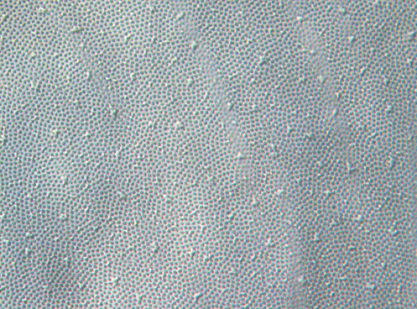
Hints: shot at 40x, it’s not part of the embryo itself, and every zebrafish pro is thoroughly familiar with it.
There was a guess that it was yolk. No! I took a quick picture of the yolk sac in this same embryo, at the same magnification.
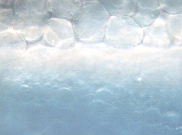
Those boulders at the top are cells, blastomeres. The bright band across the middle is the yolk syncytial layer, cells that bridge the gap between the cellular embryo and the yolk mass at the bottom. See? Nothing alike.
A few of you got it right, or came close: it’s the chorion.