Clarifying tetrapod embryogenesis, accurately
By OldCola
[Note from pzm: The text of this one is a little rougher than I like, but the content is interesting and addresses the claims of a character who has been lurking about here for a while, and whose work I’ve criticized before. If nothing else, I’d also like to see a few science posts submitted as guest articles, so think of this as priming the pump.]
The article, “Clarifying tetrapod embryogenesis, a physicistʼs point of view,” by V. Fleury, hasn’t steered the revolution expected by Fleury in evo-devo. Two years after the publication, cited by one (Fleury himself), the article seems to have being more useful to clarify the way he perceives the world, then anything related to the tetrapods embryogenesis. And the most useful elements are to be found on the Web, not in the article per se. Direct questions remain unanswered, critics are threatened by legal action for defamation, and hierarchical superiors are solicited to politely ask the critics to STFU.
While Fleury must be aware by now of major flaws in the way he represented several of the articles he used as sources of information, and of several inconsistencies of his model and the way he extrapolates his own data, he doesn’t seem to have done anything to correct them. The article remains available unchanged, a shame for EPJ AP editorial board (and Editor-in-Chief Dr DreÌvillon B. in particular), sufficiently shameful at least for the guy who invited the review, for Fleury to avoid disclosing his name.
A new element comes to complete Fleury’s quest:
V. Fleury, Dynamic topology of the cephalochordate to amniote morphological transition: A self- organized system of Russian dolls, C. R. Biologies (2011), doi:10.1016/j.crvi.2010.11.009
During evolution of vertebrates a sequence of events is empirically observed: first, animals are bilateral, but they have no heart, no head, and no surrounding bag during development (these primitive animals are called cephalochordates [1]).
From the very first phrase of the Introduction, you know hope that no biologist read the manuscript before it was accepted for publication. And certainly not any evo-devo person, which would be the right choice for a referee for this kind of subject.
Cephalochordates are certainly not vertebrates and they certainly have a head, the sub-phylum being named after the fact that the notochord extends into that head. One may think that Fleury misused the word “head”, meaning “skull” or whatever, but if you read the French summary of the paper you do get the same information, Cephalochordata don’t have a “tête” (French for “head”).
And he dare give a reference! But if you had the courage to read his previous article (for a review) you may be familiar with the strange way Fleury reports his readings (at least the way he understood them), in an absolutely surreal way, including data from his own lab! If not, there is a brand new example in this one (see below).
By the title you may have expected to read about comparative embryology/anatomy that will enlighten you on the relations between the body plans of cephalochordates and amniotes. If so, you will be deceived. Fleury focuses entirely on chicken embryos, hoping to prove experimentally the existence of some kind of order in the ontogeny of the chicken that reflects an order in the phylogeny of chordates. The reading is interesting not to learn anything about evolution or embryology (or physics by the way), but to see how an a priori can lead someone to mess up things badly. Fleury observes the world through a keyhole shaped by Plato a long time ago and he seeks some equivalent of the Holly Grail: a way to write the essence of the pattern of tetrapods without evolutionary arguments, as it “exist in the platonician space of forms“, while avoiding being embarrassed by the bullshit produced by embryologists, geneticists or evo-devo people.
The aim of this work is to support that “the formation of amniotes would be a deterministic attractor of a physical process over a flat visco-elastic plane,” and that the formation of the heart and the chorion (you should pronounce it amnios to make sense) are the consequence of the body’s growth along the anteroposterior axis.
Thus, any embryo with the amniotic (and chorionic) cavity formed before the beginning of gastrulation would falsify Fleury’s model definitively. I’ll come to that later.
While aware of the lateral folding of the embryo around an antero-posterior (AP) axis, Fleury avoid to discuss it as his model don’t explain it. Cardiac tubes are formed as mirror structures at both sides of and parallel to the AP axis, they migrate to the midline where they fuse to form the heart and they are already pre-determined to produce almost fully developed hearts if by some mutation their migration to the midline is impaired. Cardiac formation is not caused by the the cephalic fold renamed “cardiac fold” by Fleury.
The fact that the cephalic and caudal folds forming the anterior and posterior intestinal portals are distant in time by almost 24 h doesn’t bother him and his model lack any modality that would explain the latency for the formation of the posterior intestinal portal. On the contrary, he manage to represent the two folds as the result of the AP axis extension in a single schema, as being the consequences of a single phenomenon, “[f]or the sake of clarity“. He is not at his first temporal jump of embryonic structures, even of imaginary ones.
What kind of physicist could have reviewed the manuscript without requiring some kind of explanation about this particularity?
There is nothing really new in his description of the development of the chicken embryo, except the errors and omissions which make it unusable. One may prefer a classic textbook, published a while ago: Patten, B.M. (1920). The Early Embryology of the Chick. Philadelphia: P. Blakiston’s Son and Co. You can browse through it at UNSW Embryology pages, where the scans of the illustrations are of much better quality.
Some data may be interesting for people interested by the dynamics of the embryo formation, the article being based on time lapse videos of the developing embryo. There is no much of it and the graphics seem to report on single experiments (no number of observed embryos given, no variance bars on the graphics). What is really new for me, is that Fleury found a way to report a “rate of variation of the radius” of an ellipse, with a major vs minor axis ratio of ~1,5 (fig 3, a, 0′), giving a single value! Any mathematician around to explain us this?
As Fleury decided to rename the formation of the subcephalic pocket “cardiac fold”, and he was seeking some symmetry at the caudal region, he also renamed the subcaudal pocket “cardiac fold” and he triumphantly mention the “aneural heart” of the hagfish as an evidence of the power of prediction of his model. Now, the caudal heart of the hagfish is just a pair of specialized structures on the caudal veins, parallel to the AP axis, as the primitive heart tubes, separated by a cartilage septum and they are innervated! Jensen, in the Introduction of his paper clearly explain the anatomy of the circulatory system of the hagfish and what elements are innervated, or not. Either Fleury didn’t bothered reading the paper or he is simply unable to understand what he is reading (or both, your guess). It would have be nice if he had read the paper, because he passed over the existence of the portal heart and of what some people call the cephalic hearts of the hagfish (specialized gill musculature which propel the blood through the arterial circulation). There is even an illustration for people bored by textual explications (fig 4). Such a little animal, so many hearts and not enough folds to explain them. Unnerving.
Patten starts his Introduction by a very wise advice:
The only method of attaining a comprehensive understanding of embryological processes is through the study and comparison of development in various animals.
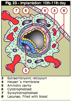
As I said, any embryo with the amniotic cavity formed before the beginning of gastrulation would falsify Fleury’s model definitively. Let me present you an artist’s rendition of Dr Fleury at his early youth, second week of development
.
The illustration is from the online Human Embryology course notes (clic the image for the full page). I’m not sure they had in mind Vincent Fleury when they draw this cartoon, but it’s the best I can offer you: A cute embryo with his amniotic cavity lined with cells from the epiblast and his primary yolk sac lined by cells derived by the hypoblast. The Heuser’s membrane is still attached to the extraembryonic reticulum.
A few days later, the secondary yolk sac had formed and the chorionic cavity was installed.
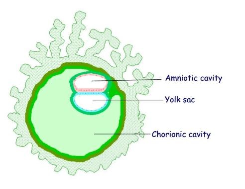
At this stage little Vincent was still bilaminar with fully formed amniotic and chorinic cavities.
Those of you interested to learn about embryo’s folding can also visit Folding of the germinal disk and the generation of the abdominal wall, in which case the comparison of the two foldings (cephalo-caudal and lateral), animation is a must for the visitor, and certainly for Fleury.
How sad that a great model from an experimentalist working all day with embryos, goes down the drain after being confronted to elements of chapter 5 of a Human Embryology textbook.
Several questions come in mind in this situation, the first one being: who the hell reviewed the manuscript. Not a biologist, probably not a physicist (he should have ask for a mechanism explaining the delay of formation of the caudal fold). Not a second year student of biology or medicine neither; she would have spotted the problem with the amniotic and chorionic cavities subito presto.
Fleury’s precedent paper was an invited review by one of the editors of a journal of physics. You can’t blame the guy for being unable to understand the bunch of errors the review contains. OK, you can blame him for not having a specialist’s opinion on the final piece of work. Misplaced trust. And sometimes, some physicists are just pissed-off by life scientists. Fleury didn’t even dared to give his name.
This time, the journal is a publication of the French National Academy of Science and it displays “Biologies” on the cover. Shame on them. Until this paper is retracted who would trust the “Development and reproduction biology” section of the journal, or the journal at all? I wouldn’t, would you?
“Therefore, this suggest” is one fabulous transition.
The Methods section of the paper may be interesting if you plan a few experiments with chicken embryos, but dramatically incomplete. The most interesting part is missing: the references of the software and the method Fleury is using for PIV, which gives him astonishing images. I would like to be able to check by myself, previous interpretations of experimental data, even the ones generated in his lab, by Fleury being as much surreal as his usual stuff. Hopefully he could complete this section in the comments of this post.
In Heart formation, Fleury undergo to explain how the heart is formed by the heart fold. Here is the first part where it goes really bad. I can understand the frustration of a physicist who would like to have more data concerning the biomechanics of the process, and hopefully somebody else than Fleury will go for them. But there is no need to reinvent the wheel, there are nice descriptions of the movements by which the heart tubes are forming, how the lateral folding of the embryo make them join along the anteroposterior axis and describing their fusion to produce the unique heart tube [1]. Certainly, the 125° rotation of the heart fields and the lateral folding of the embryo necessary for the normal cardiogenenic process are not perpendicular to the anteroposterior axis and doesn’t fit Fleury’s model, but it isn’t reasonable to just ignore them. You can’t just ignore what it doesn’t fit your model to make it sound plausible.
Anyway, even the fusion of the primary heart tubes doesn’t seem to be necessary to support the development and morphogenesis of the heart, up to some point: “a highly differentiated four-chambered mammalian heart” in the case of Foxp4 mutant mice embryos [2].
The point of junction of the cardiac tubes do travel caudaly along the anteroposterior axis of the embryo, but that’s just the point of junction…
An interesting description of the heart formation can be found in a relatively old textbook: The Early Embryology of the Chick (pp 68-72, fig. 26 & 27, with emphasis for fig 27) [3]
For those who will take the time to read the paper, please pay attention to the part discussing the role of chemotactic forces ; Fleury didn’t managed yet to understand morphogenic gradients and that most of them are embedded into the cells and the extracellular matrix.
You may need to go through the whole section about the Chorion formation to understand that Fleury discuss just about the amniotic folds of the chorion and completely ignores the rest of it. It’s just that it isn’t folded in the right direction for his model. On the other hand the amniotic folds of the chorion are folded in the right way and Fleury carefully studied the ways the meet around a single point. Not only it’s weird how he doesn’t discuss the lateral part of the amniotic folds (absolutely necessary to form the amnios and the dorsal part of the chorion), but not perpendicular to the anteroposterior axis, but somehow he manage to found a single rate of variation of the radius of an ellipse!
Patten [3] offers a series of diagrams showing the growth and foldings of the somatopleure which form the amnios, from transverse sections of the embryo, in fig 30 and from longitudinal sections in fig. 32. That gives a global image of the tissue growth, in all directions, not just the keyhole presentation Fleury is giving in his article.
While Fleury is aware that the cephalic and caudal amniotic folds appear at different developmental stages, he present their occurrence as being caused by the “extension of the median axis” without explaining what may be the mechanical causes for the delay of almost 24h for the apparition of the caudal amniotic fold. “For the sake of clarity” he present them in the same figure (4b of his paper) as if they occurred in the same time. As much clarity as usually.
1. Heart Field: From Mesoderm to Heart Tube, Radwan Abu-Issa, and Margaret L. Kirby, Annual Review of Cell and Developmental Biology Vol. 23 (2007): 45-68, doi: 10.1146/annurev.cellbio.23.090506.123331
2. Advanced Cardiac Morphogenesis Does Not Require Heart Tube Fusion, Shanru Li, Deying Zhou, Min Min Lu, Edward E. Morrisey, Science Vol 305 (2004): 1619-1622, doi: 10.1126/science.1098674
3. Patten, B.M. (1920). The Early Embryology of the Chick [link to scans in pdf at archive.org]. Philadelphia: P. Blakiston’s Son and Co. You can browse through it at UNSW Embryology pages, where the scans of the illustrations are of much better quality.
V. Fleury, Dynamic topology of the cephalochordate to amniote morphological transition: A self-organized system of Russian dolls, C. R. Biologies (2011), doi:10.1016/j.crvi.2010.11.009
