Don’t you just love those photo series of the young’uns at different ages?
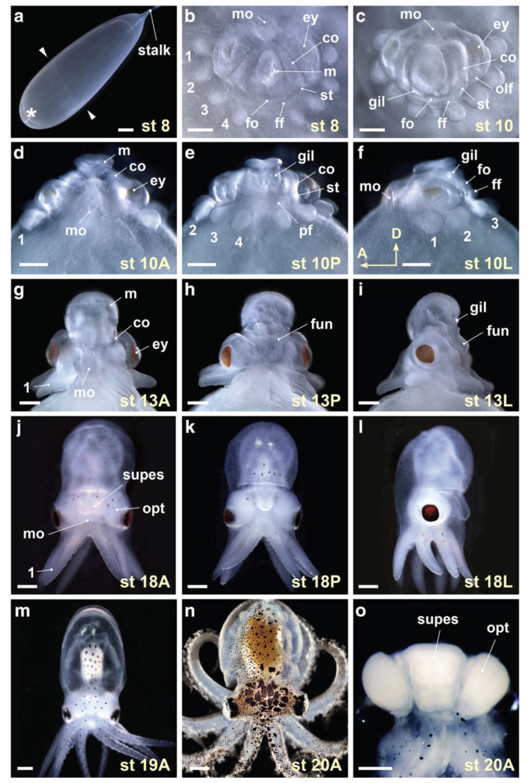
Developmental staging of Octopus bimaculoides. a Whole egg photomicrograph illustrates the egg stalk and the animal pole (asterisk) where the embryonic body forms. Extent of epiboly in this stage (st) 8 embryo is marked with arrowheads. b End on view of a stage 8 embryo with the egg capsule and yolk removed. In dark field illumination, the organ primordia are visible as ectodermal and mesodermal thickenings. The mantle anlage (m) is central, the prospective mouth (mo) at the top of the panel is anterior, and the arm bud pairs (1–4) are arrayed peripherally. The folds of the collar (co) and the prospective funnel (fo, ff) fall at intermediate positions. c–f The growth of the organ systems by stage 10 is illustrated in end on (c), anterior (d), posterior (e) and left side (f) views. g–m The shape of the adult octopus emerges at middle (g–l) and late (m) embryonic stages. Illustrated are anterior (g and j), posterior (h and k) and left side (i and l) views of stage 13 (g–i) and stage 18 (j–l) embryos, and an anterior view of a stage 19 embryo (m). n and o Anterior views of O. bimaculoides (n) and its brain (o) at hatching (stage 20). A, anterior view; ey, eye; fun, funnel; gil, gill; L, lateral view; olf, olfactory organ; opt, optic lobe; P, posterior view; pf, funnel pouch; st, statocyst; supes, supraesophageal mass. Scale bars: 1mm (a), 500 μm (b–o)
Shigeno et al. Zoological Letters (2015) 1:26

Interesting, during the developmental stage depicted in the 8th photo, they appear to be made entirely of fun.
I can honestly say that I’ve never wanted an embryo as a pet before.
It’s fun watching it turn into an octopus!
And kind of adorable.